Lipid Identification: Chemical Fingerprint Instead of Dye
A team at Helmholtz Munich and the Technical University of Munich (TUM) has developed a new microscopy technique that can distinguish lipid species in living cells – in particular cholesterol and sphingomyelin – and map them without the need for chemical labeling. By combining mid-infrared illumination with optoacoustic detection, the method reads the lipids’ natural spectral fingerprints, eliminating the need for specific fluorescent tags, which are laborious to develop and may interfere with lipid function. The team reports its results in the journal Nature Methods.
Lipids are key building blocks of cell membranes and help control how cells transmit signals and transport substances. However, sensing or visualizing specific lipid classes in living cells is challenging. Traditional fluorescence microscopy requires developing custom fluorescent labels for each lipid – a time-consuming and costly process – and these labels can sometimes affect the lipid’s function, stress the cells, or fail to bind specifically to the intended target.
Mid-Infrared Light and Ultrasound Make Lipids Visible Without Labels
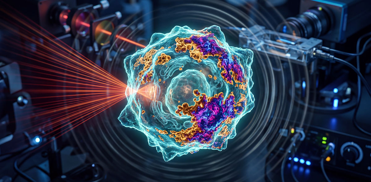
A team led by Prof. Vasilis Ntziachristos, Head of the Bioengineering Center and Director of the Institute of Biological and Medical Imaging (IBMI) at Helmholtz Munich, as well as Professor and Director of the Chair of Biological Imaging at the Technical University of Munich, has tackled this challenge by developing a new microscopy method called hyperspectral mid-infrared optoacoustic microscopy (HyFOPM), which makes lipids visible without labels.
The method illuminates the sample with pulsed mid-infrared light at multiple distinct wavelengths, a so-called “hyperspectral” illumination. Lipids absorb specific bands of this light, producing a tiny, brief temperature rise that generates ultrasound waves. These waves are detected by ultrasound transducers and converted into a spectral image. Computational analysis of this spectral image then produces maps showing the distribution of different lipids within the sample.
“The absorption pattern is characteristic of different molecules and acts like a unique molecular fingerprint,” says Dr. Francesca Gasparin, a scientist at IBMI and TUM, and first author of the study. “This mid-infrared fingerprint allows us to distinguish different lipids without requiring external labels”.
The Fingerprint Region Makes the Difference
What makes this method unique is the wavelengths it uses. Many label-free approaches rely on spectral ranges dominated by bonds found in a wide variety of biomolecules, producing similar signals that make it hard to distinguish individual lipid classes.
The team instead focuses on the so-called fingerprint region, where absorption features arise predominantly from vibrational modes that are highly characteristic to each lipid’s molecular structure. This means the method can detect not just the building blocks of a molecule, but also how they are arranged in relation to each-other. By operating in this region, the technique can differentiate between lipid species – even chemically similar ones such as glycerophospholipids and sphingomyelin.
Minimal Stress for Cells
To test the reliability of these chemical fingerprints, the team compared their measurements with conventional infrared spectroscopy, which served as a reference. While the traditional method provides accurate measurements of dissolved compounds and solution droplets, it is not suitable for living cells, which require special preparation that can stress the cells.
The new microscopy technique not only reproduced the expected lipid spectra in solution but also enabled measurements directly in living cells. “A key advantage of HyFOPM is that label-free lipid observation results in minimal stress for living cells,” says Francesca Gasparin.
Outlook: From Cell Culture to Patient-Adjacent Applications
In the long term, the researchers see applications across a wide range of lipid research, from basic science to medical applications that have so far been difficult to access.
“Being able to track and map lipid classes in living cells without labels opens new ways to understand disease processes and monitor metabolic activity – not only in cells but also in humans,” says Prof. Vasilis Ntziachristos. “The next step is to speed up the method and test it systematically in complex cellular systems, to build knowledge on disease development and drug action. Ultimately, we aim to use it in humans for continuous monitoring of metabolites, providing actionable biomarkers across a wide range of conditions within the cardiometabolic syndrome and beyond.”
Original publication
Francesca Gasparin, Alexander Prebeck, Alice Soldà, Nasire Uluç, Sarah Glasl, Constantin Berger, Miguel A. Pleitez, Vasilis Ntziachristos; "Differentiation of sphingomyelin and cholesterol by hyperspectral mid-infrared detection of single-bond vibrational modes in the fingerprint region"; Nature Methods, 2026-3-30

Other news from the department science


Most read news


More news from our other portals


See the theme worlds for related content
Topic World Spectroscopy
Investigation with spectroscopy gives us unique insights into the composition and structure of materials. From UV-Vis spectroscopy to infrared and Raman spectroscopy to fluorescence and atomic absorption spectroscopy, spectroscopy offers us a wide range of analytical techniques to precisely characterize substances. Immerse yourself in the fascinating world of spectroscopy!
Topic world Fluorescence microscopy
Fluorescence microscopy has revolutionized life sciences, biotechnology and pharmaceuticals. With its ability to visualize specific molecules and structures in cells and tissues through fluorescent markers, it offers unique insights at the molecular and cellular level. With its high sensitivity and resolution, fluorescence microscopy facilitates the understanding of complex biological processes and drives innovation in therapy and diagnostics.