Revolutionizing cancer research with diamonds
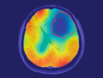
magnetic resonance imaging (MRI) is being revolutionized: With the nanodiamond the tumor tissue can be detected sooner and distinguished better from the healthy surrounding tissue. Aiming at an improvement of the MRI procedure in the joint project »DiaPol« Fraunhofer IAF cooperates with the University of Ulm, the company NVision imaging Technologies GmbH, the Hebrew University of Jerusalem and the Israeli Center for Advanced Diamond Technologies (ICDAT). The novel technology offers great opportunities: the extremely precise and quickly available results make it possible to adjust the treatment of the tumor tissue to the patient in a significantly more efficient way than it has ever been possible with previous methods.
It’s the uncertainty that frightens us most: Cancer. According to a report by the Robert Koch Institute from 2016, the absolute number of new cases in Germany has almost doubled since the early 1970s. Time is a crucial factor, as an early and accurate diagnosis can save lives. During the past decades the methods to detect suspicious tissue in the body have continuously become more precise. Magnetic resonance imaging is particularly gentle and efficient for patients because it works without any harmful chemicals or radioactive substances. MRI can also create three-dimensional, detailed cross-sections of the human tissue.
Classical MRI uses magnetic fields in order to produce high-resolution images. A human body is composed of 70 percent water. Each water molecule contains two hydrogen atoms with magnetic nuclei. The magnetic fields in these nuclei are generated by nuclear spins. A so-called polarisator can be used in order to amplify and adjust the tiny magnetic fields of these spins. The better the spins are adjusted, the stronger is the MRI’s signal and the more accurate the results. By adding high frequency pulses, certain atomic nuclei in the human body are excited resonantly, which can be measured as an electrical signal. A program subsequently translates the signals into high-resolution, three-dimensional images.
10,000 times more sensitive thanks to diamond-based polarizers
For the novel MRI procedure, the researchers combine the classical method with a nanodiamond polarisator. Built-in nitrogen vacancy centers in a diamond play an important role in the polarizer of the innovative process: the electron spins in these centers generate magnetic fields that can be transmitted to other nuclear spins, and thus adjust them (»polarize« them). This procedure hyperpolarizes the nanodiamonds or external molecules. They can then be injected into the human body before the MRI scan, which significantly increases the imaging sensitivity. As an expert in the field of diamond nanotechnology, Fraunhofer IAF is involved in this part of the project.
»Our tasks are the diamond’s optimization on the nanoscale and the incorporation of the nitrogen vacancy centers«, explains Dr. Verena Zürbig from Fraunhofer IAF. The project coordinator and group leader for »Diamond Technology« is convinced: »Compared to the conventional procedure, the diamond polarizers will significantly increase the MRI’s sensitivity.« The company NVision sees great prospect in the new procedure: »Not only could it become possible to diagnose cancer early, but also to identify the cancer cell’s exact stage.«
Diamond as a material has some unbeatable advantages. For example, the hyperpolarization with diamond can be achieved at room temperature, thus enabling a much faster and cost-efficient method in comparison to conventional procedures, which still require very low temperatures. One of the project’s sub-goals is the construction of extremely small, flexible and mobile diamond polarizers. This innovation enables fast analyzing and shortens the time for patients waiting for their results from several weeks to a few days. By providing more precise measurements and a corresponding improved treatment, the project hopes to bring relief to patients often struggling with the uncertainty and fear caused by cancer.
Most read news
Topics
Organizations
Other news from the department science

Get the analytics and lab tech industry in your inbox
By submitting this form you agree that LUMITOS AG will send you the newsletter(s) selected above by email. Your data will not be passed on to third parties. Your data will be stored and processed in accordance with our data protection regulations. LUMITOS may contact you by email for the purpose of advertising or market and opinion surveys. You can revoke your consent at any time without giving reasons to LUMITOS AG, Ernst-Augustin-Str. 2, 12489 Berlin, Germany or by e-mail at revoke@lumitos.com with effect for the future. In addition, each email contains a link to unsubscribe from the corresponding newsletter.


