Discovered by chance: the refractive index microscope
An astonishing success: by combining completely different microscopy methods, the optical density of a sample can be measured with pinpoint accuracy

The original intention was to examine biological samples on a molecular scale and encountered stubborn problems. But then it was discovered that the cause of the annoying measurement inaccuracy, the variable refractive index of the sample, can be precisely determined and thus becomes a highly interesting measurement result itself - if two fundamentally completely different microscopy methods are combined.
Almost by chance, TU Wien succeeded in developing a new type of microscopy technique that can be used to measure the refractive index of biological samples - with a resolution far below what light microscopy should allow according to conventional theory.
The trick for sub-wavelength resolution
What happens if you want to photograph two molecules whose distance is smaller than the wavelength of the light? Then you won't see two separate dots, but a single blob of light - the images of the two molecules overlap, no matter how precisely the microscope works.
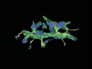
But there is a way out - so-called "single-molecule microscopy". Special molecules are incorporated into the sample that flash at different times. Each of them creates a small disc of light in the camera, and if you measure the center of this disc, you know exactly where the molecule is located. Even if there is another molecule within the same disk - if they light up one after the other and can be measured separately, they can both be imaged precisely. While their images would simply blur together in an ordinary microscope image, this method allows extremely high-resolution images.
"However, the light discs that are measured in this way are not always the same size," says Prof. Gerhard Schütz from the Institute of Applied Physics at TU Wien. "The size of the light disk depends, for example, on how close the molecule is to the focal plane of the camera." The original intention was to use precisely this phenomenon as a useful source of information: If the distance of the molecule could be determined from the size of the light disks, then a 3D image could theoretically be generated from the light disks. But it soon became clear that it is not quite that simple.
Does the distance change - or the refractive index?
"The problem is that the size of the light disks also depends on the refractive index of the material," explains Gerhard Schütz. Not every material allows light rays to pass through at the same speed, and it is precisely this effect that causes light to be deflected by prisms or lenses. There are therefore two parameters that can have an influence on the measured light spot - distance and refractive index.
But what if you make a virtue out of this very necessity? "We decided to simply reverse this problem," says Gerhard Schütz. "We simply measure the 3D structure of our sample in a different way, namely with an atomic force microscope. We can then use our light image to calculate the refractive index at every point of our sample with pinpoint accuracy."
New measurement method for biological materials research
In cooperation with the Medical University of Innsbruck, the team at TU Wien has developed a technique that can be used to measure the refractive index of biological samples on a scale well below the wavelength of light.
"This is particularly exciting when it comes to collagen in tissue," says Gerhard Schütz. "Collagen can absorb different amounts of water, and the refractive index changes accordingly. With our method, we can now determine exactly how much water is in which place. We can also obtain data on the chemical composition of the tissue that was previously not directly accessible." The result - triggered by an almost accidental discovery - is a new link between physical measurement technology and microbiological research.
The research was funded by the FWF and the WWTF and resulted from a collaboration between the Institute of Applied Physics and the Institute of Lightweight Structures and Structural Biomechanics at TU Wien and the Institute of Biomedical Physics at the Medical University of Innsbruck.
Note: This article has been translated using a computer system without human intervention. LUMITOS offers these automatic translations to present a wider range of current news. Since this article has been translated with automatic translation, it is possible that it contains errors in vocabulary, syntax or grammar. The original article in German can be found here.
Original publication
Simon Jaritz, Lukas Velas, Anna Gaugutz, Manuel Rufin, Philipp J. Thurner, Orestis G. Andriotis, Julian G. Maloberti, Simon Moser, Alexander Jesacher, Gerhard J. Schütz; "Refractive Index Mapping below the Diffraction Limit via Single Molecule Localization Microscopy"; ACS Nano, Volume 20, 2025-12-26

Other news from the department science


