Nanomaterial self-assembly imaged in real time
A team of researchers from UC San Diego, Florida State University and Pacific Northwest National Laboratories has for the first time visualized the growth of 'nanoscale' chemical complexes in real time, demonstrating that processes in liquids at the scale of one-billionth of a meter can be documented as they happen.
The achievement, which will make possible many future advances in nanotechnology, is detailed in a paper published in the Journal of the American Chemical Society. Chemists and material scientists will be able to use this new development in their basic and applied research, for example, to better understand the stepwise formation of nanostructures.
Previously, scientists could examine changes in nanostructures only by looking at the large-scale alterations of a bulk population of particles or by taking 'screen shots' in a static fashion of individual nanostructures with electron microscopy.
'That process is like taking photos every 10 minutes of a football game and then trying to piece these photos together to tell the story of what is really a highly dynamic process,' said Nathan Gianneschi, an associate professor of chemistry and biochemistry at UC San Diego who headed the research effort with Seth Cohen, chair of UC San Diego's Department of Chemistry and Biochemistry.
'Until now, this was the state of the art in terms of how we could document how nanostructures formed. The development we describe in our paper demonstrates that these processes can be observed in real time, by literally videoing these processes on the nanoscale level using an electron microscope.'
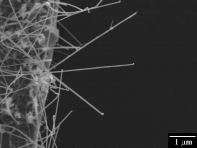
The development employed a recently developed process called Liquid Cell Transmission Electron Microscopy. Transmission Electron Microscopy, or TEM, has long been used by scientists to image nanoscale materials and understand nanoscale structure. While advances in Liquid Cell TEM, or LCTEM, had permitted scientists to visualize the motion of nanoscale objects in liquids, researchers had not yet figured out a way to use it to visualize the growth of complex self-assembled, chemical nanostructures.
'We showed for the first time that this technique can be used to observe the growth of complex organic-inorganic hybrid materials, providing an unprecedented understanding of their formation,' said Gianneschi. 'This demonstration marks a significant step forward in LCTEM becoming essential for our understanding of nanoscale processes for all materials in liquids.'
The team of scientists included Joseph Patterson and Michael Denny of UC San Diego, Patricia Abellan, Nigel Browning and James Evans of Pacific Northwest National Laboratory and Chiwoo Park of Florida State. Patterson, the first author of the paper, did all of the Liquid Cell Transmission Microscopy at instruments at UC San Diego and PNNL with the assistance of Evans, who is an expert in the technique, while Park was responsible for the video analysis.
To make things simple, the researchers initially set out to study a chemical system known to assemble with a limited number of components and give rise to well-defined materials.
'We considered metal-organic frameworks to be the perfect starting point for this because they give ordered structures through an assembly process and include organic and inorganic components,' said Gianneschi. 'The first step was to determine if these nanostructures would survive the experiment. This is necessary because materials are susceptible to being destroyed by the high energy electron beam that is used to image them. Once these conditions were established, we were then able to flow components into the TEM instrument, in solvent, and watch as the assembly process took place. This was made possible using a special sample cell holder for the TEM that allowed us to put liquids within a chamber, within the high vacuum instrument. We could then image through the chamber, to see what's inside.'
The scientists' demonstration that such chemical complexes can be imaged in real time suggests that the complex processes of other 'delicate self-assemblies' could be elucidated in greater detail, such as biologically produced chemicals and viruses, which are more than a thousand times smaller than bacteria.
'This advance provides a tool for observing material as they assemble with resolutions only possible using electron microscopy,' Gianneschi said. 'That is, length scales can be observed that are relevant to nanoscale materials and processes. In terms of imaging dynamics like this, we believe it will impact how nanotechnology is developed in the future.'
Other news from the department science


