F is for Fluoresence and Fluorine
New dyes for optical nanoscopy
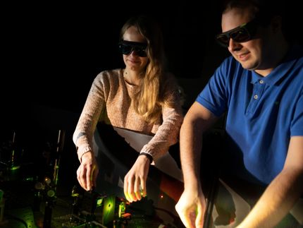
The imaging of living cells at the molecular level was barely a dream twenty years ago. Today, however, this dream is close to becoming reality. In the Max Planck Institute for NanoBiophotonics in Göttingen, Stefan Hell has developed fluorescence microscopy methods for observing objects on the nanoscale and with his colleagues Vladimir Belov and Christian Eggeling a new series of photostable dyes that can be used as fluorescent markers has been realised, as reported in a cover story in Chemistry—A European Journal.
Over the last two decades Stefan Hell and his group have revolutionized the art of microscopy beyond limits thought to have been unbreakable. Due to the wave properties (diffraction) of light, the resolution of an optical microscope is limited to object details of about 0.2 micrometers. The laws of physics appeared to prohibit imaging details beyond this limit. Stefan Hell saw beyond this limitation and about fifteen years ago his vision became concrete; he developed a method for observing objects at the nanometer scale by sequentially turning the fluorescence of nearby molecules off by stimulated emission, a technique known as STED nanoscopy.
The sensitivity of this technique depends on the brightness of the applied fluorescence markers and their photostability is also of great importance. The NanoBiophotonics group has succeeded in synthesizing a series of highly photostable and highly fluorescent dyes. These compounds emit green and orange light and are based on fluorine derivatives of the well-known Rhodamine dye. The use of these dyes in STED nanoscopy leads to images of high-quality with respect to brightness and signal-to-background ratio; further the resolution over that of more traditional optical microscopes is significantly improved giving more detailed structural information.
These rhodamine-based fluorine derivatives are even more special because of their versatility. The compounds are available in hydrophilic and lipophilic forms, and with the inclusion of amino reactive groups, they can be easily attached to antibodies or other biomolecules in the course of standard labeling and immunostaining procedures. The group demonstrate that these new dyes are able to cross cellular membranes and reach the interior of living cells, which could lead to new labeling strategies for biological systems. All eyes are now on Göttingen to see just how far optical nanoscopy can go.
Original publication: Stefan Hell et al.; "New Fluorinated Rhodamines for Optical Microscopy and Nanoscopy"; Chemistry - A European Journal 2010, 16, No. 15, 4477–4488.
Other news from the department science


These products might interest you


Most read news


More news from our other portals


See the theme worlds for related content
Topic world Antibodies
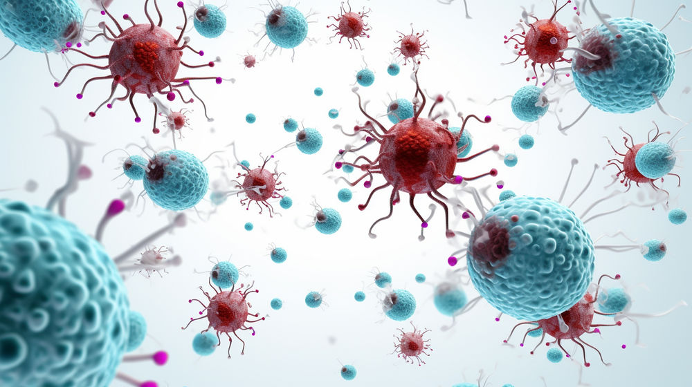
Antibodies are specialized molecules of our immune system that can specifically recognize and neutralize pathogens or foreign substances. Antibody research in biotech and pharma has recognized this natural defense potential and is working intensively to make it therapeutically useful. From monoclonal antibodies used against cancer or autoimmune diseases to antibody-drug conjugates that specifically transport drugs to disease cells - the possibilities are enormous
Topic world Antibodies
Antibodies are specialized molecules of our immune system that can specifically recognize and neutralize pathogens or foreign substances. Antibody research in biotech and pharma has recognized this natural defense potential and is working intensively to make it therapeutically useful. From monoclonal antibodies used against cancer or autoimmune diseases to antibody-drug conjugates that specifically transport drugs to disease cells - the possibilities are enormous