Revealing Photographs of DNA
Ultrasensitive DNA detection with photographic paper
Ultrasensitive genetic detection methods could revolutionize the diagnosis and treatment of diseases. However, all the techniques until now have been far too technically demanding for broad application. Munich researchers led by Thomas Carell have now developed a very simple method based on the intensification process used in black-and-white photography. As reported in the journal Angewandte Chemie, this method should in principle be able to detect DNA at attomole (10-18 mol) levels.
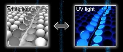
In black-and-white photography, light is absorbed by silver bromide crystals in a light-sensitive layer. This forms little heaps of silver atoms. In the subsequent development process, these atoms then catalyze the reduction of the silver ions of the entire crystal to elemental silver, which turns the photographic paper black in the areas exposed to light. In principle, this process can reach an intensification factor up to a hundred billion (1011).
Common photographic paper only reacts to UV and blue light, but not to red light. To also capture red objects, a photographic sensitizer must be added to the photographic paper. This is what the researchers have used to "photograph" DNA: They attached this type of sensitizer to the DNA fragments to be detected. Under darkroom conditions, they sprinkled the DNA solution onto red-sensitive photographic paper and irradiated it with red light. Once the paper was developed, black spots visible with the naked eye appeared wherever the DNA solution came into contact with the paper.
To prove that pathogens can also be revealed photographically, the team selected a strand of DNA that has at its center a segment complementary to a short sequence in the genetic code of the pathogen that causes the plague. The two complementary segments at the ends of the strand bind together and force the strand into a hairpin shape. The researchers attached a fluorescent dye to one end of this "probe" and a fluorescence quencher to the other end. When in the hairpin form, the dye and quencher are close to each other, which shuts off the fluorescence. If plague DNA is present in the sample, it binds to the center segment of the probe, which opens the hairpin and causes the dye and quencher to move apart from each other-the fluorescence is "switched on" and the photographic paper is sensitized. When sprinkled onto photographic paper and irradiated with light, samples containing the probe always generate black spots when plague DNA is present in the sample.
Original publication: Thomas Carell et al.; "DNA Photography: An Ultrasensitive DNA-Detection Method Based on Photographic Techniques"; Angewandte Chemie International Edition 2007.
Other news from the department science


Most read news


More news from our other portals


Last viewed contents
Assessing quality of flowing waters with DNA analyses - Organisms in streams and rivers shed light on quality of waters
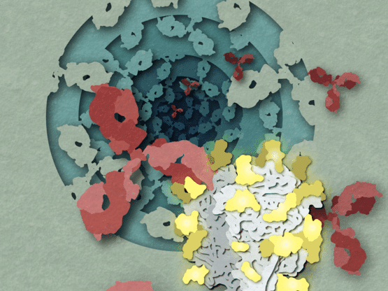
Deadly virus structures point toward new avenues for vaccine design - By comparing the structures of protein complexes from different lineages of the dangerous Lassa virus, a Scripps Research team identified new antibodies and vaccine targets
Final report: analytica Anacon India shows increase in all areas