The need for speed
Coherent Raman scattering methods have one key advantage over spontaneous Raman microscopy: speed. The (sub-)microsecond pixel dwell times offered by narrowband CRS imaging methods have initiated a new era of chemical imaging applications in biology and biomedicine.
Biomedical scientists are dreaming of a technique showing the distribution of all the biomolecular constituents that make up biological tissue in high-resolution, three-dimensional maps. Such a visualization tool does not currently exist. Whereas fluorescence and refraction based techniques will never be able to identify an arbitrary molecular compound in tissue, vibrational imaging techniques offer much more promise. They work label-free and non-invasively and they are able to identify many important groups.
The conventional vibrational imaging technique is Raman microscopy. Practical limitations have so far prevented Raman microscopy from reaching its full potential. The most important limitation is speed. The intrinsically weak Raman signals severely limit the achievable image acquisition rate. The development of coherent Raman scattering (CRS) microscopy techniques over the last decade has resulted in important steps toward resolving the speed issue. In a feature article, Eric O. Potma and a team of scientists at the University of California (Irvine, USA) discuss several ways in which the improved speed of CRS microscopy has transformed the biomedical imaging field and touch on some of the challenges that lie ahead in moving towards the realization of a more generally applicable visualization technique.
Stronger signals are obtained because in CRS the molecules are driven coherently, which makes them radiate in unison. The resulting signal is coherently amplified through constructive interference in a well-defined, phase-matched direction which enables efficient detection of the Raman response. For instance, when the microscopic focal volume is filled with lipids, the number of detected photons in coherent anti-Stokes Raman scattering (CARS), generated from the CH2 stretching mode, can easily exceed 102 per microsecond at 10 mW of illumination. With such high signal levels, real-time Raman imaging of biological tissues becomes feasible.
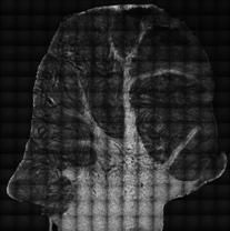
An important application of high speed CRS imaging is the visualization of large tissue segments. For example, neural injuries and myelination disorders in live spinal tissues have been mapped as well as atherosclerotic plaques in aortas. Tissue maps were generated that cover up to several centimeters in lateral and hundreds of microns in axial distance, while preserving the (sub-)micrometer resolution offered by the high numerical aperture objective.
The intrinsic movement of the tissue, due to pulsation of blood vessels, breathing, or positional adjustments by the subject, poses a challenge when imaging living tissues. Imaging at video-rate enables image acquisition in which the individual frames show minimal blurring due to movements. Another advantage of fast imaging is the reduction of possible photodamage in living tissues. Besides live tissue imaging in small animal models, CRS imaging has already been applied to the examination of human skin in vivo.
In addition, the fast imaging modalities enable a time- and space-resolved view of dynamic processes of biological relevance. Water diffusion in neutrophile cells, the dynamics of intracellular droplets, and the diffusion of pharmaceutically relevant agents through skin are examples of dynamic processes that have been visualized.
Single frequency CARS and SRS have already proven indispensable in the study of lipids and lipid metabolism in live tissues and cells. Because of its imaging speed and chemical contrast, CRS has pushed the concept of label-free and noninvasive chemical imaging closer to clinical biomedical applications. The challenge ahead is to marry the speed qualities of single frequency scanning with the superior spectral information of broadband Raman spectroscopy. Recent developments suggest several approaches by which this could occur and provide a glimpse toward the ideal of clinically relevant, real-time chemical inspection of live tissues.
Original publication
J.L. Suhalim et al.; J. Biophotonics 5, 387-395 (2012)
Other news from the department science


