Skin Cancer on the Face: Ultrasound Shows How Deep the Tumor Grows
Summer, sun, UV radiation: The warm season puts particular strain on the skin—especially on the face. The nose, ears, forehead, and scalp are often exposed to the sun without protection for years on end. It is precisely in these areas that skin cancers such as basal cell carcinoma and squamous cell carcinoma frequently develop. When such tumors must be surgically removed, it’s not just what’s visible on the surface that matters: how deep the tumor has already grown is just as important. High-frequency ultrasound can help provide a better answer to this question before surgery for certain skin tumors, explains the German Society for Ultrasound in Medicine (DEGUM).
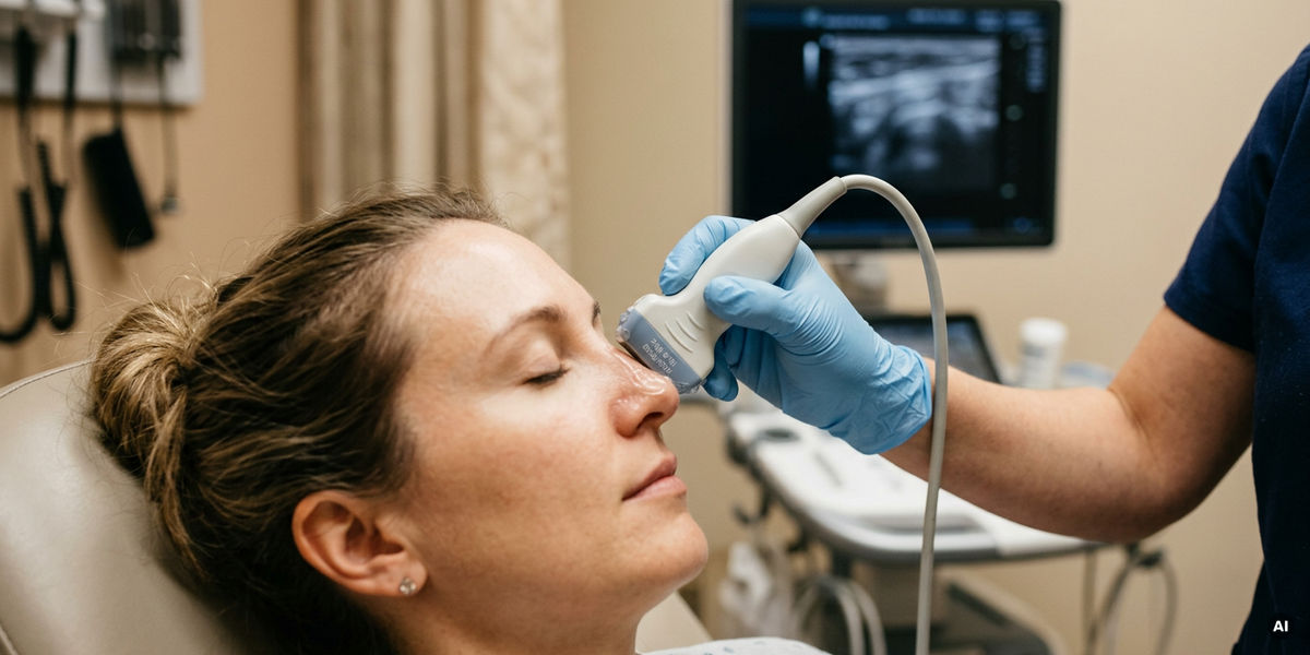
Many people associate ultrasound primarily with pregnancy or examinations of abdominal organs. Yet sonography has long been indispensable in many medical specialties—including dermatology. In the case of skin tumors, it provides a view of the tissue beneath the visible lesion. This is particularly relevant, for example, on the nose, where skin, fatty tissue, cartilage, and bone are located in close proximity to one another.
Even if a skin lesion appears small on the surface, surgery sometimes requires careful planning—for example, if the tumor has grown deeper toward the cartilage. If this extent only becomes apparent during the procedure, it complicates the removal of the tumor and the subsequent reconstruction. On the nose in particular, this can have consequences for appearance and, in some cases, for breathing as well. “With high-frequency ultrasound, we can better assess before surgery whether a tumor is growing only superficially or whether adjacent structures are affected,” explains Dr. Diana Crisan, MD, senior physician at the Department of Dermatology and Allergology at Ulm University Hospital and a member of the DEGUM Dermatology Section. “This helps us provide patients with more realistic advice and consider potential reconstruction at an early stage.”
How Ultrasound Influences Treatment Planning
During the examination, a special ultrasound probe is used that can very accurately visualize the tissue layers directly beneath the skin. The findings can influence which treatment options are considered: For very superficial basal cell carcinomas, local treatment methods such as creams, light therapy, or cryotherapy may be possible under certain conditions. If, on the other hand, the ultrasound shows that a tumor extends deeper or is growing irregularly, this tends to indicate a need for surgery, which must be planned with appropriate care. In cases where findings suggest advanced disease progression—for example, damage to the underlying bone that can be clearly visualized on ultrasound—surgery is generally not recommended. In such cases, other forms of treatment may be considered, such as modern medications or radiation therapy. “Laboratory examination of the tissue also remains important,” Crisan emphasizes. “This helps confirm the diagnosis and verify whether the tumor has been completely removed.”
Preoperative Ultrasound: Pain-Free and Radiation-Free
Ultrasound is painless, radiation-free, and usually available quickly. For skin tumors close to the surface, the examination can be integrated directly into dermatological diagnostics. In select cases, ultrasound can also be used to monitor whether local therapies are effective.
High-frequency ultrasound is not yet standard practice everywhere in skin cancer surgery. The method requires experience, a trained eye, and expertise in dermatology and oncology. The DEGUM is therefore committed to training based on established quality standards and to the broader integration of modern ultrasound procedures into patient care.
Note: This article has been translated using a computer system without human intervention. LUMITOS offers these automatic translations to present a wider range of current news. Since this article has been translated with automatic translation, it is possible that it contains errors in vocabulary, syntax or grammar. The original article in German can be found here.
Original publication
Ximena Wortsman; "Ultrasound in Skin Cancer: Why, How, and When to Use It?"; Cancers, Volume 16, 2024-9-27
Diana Crisan, Ximena Wortsman, Orlando Catalano, et al.; "Pre‐operative high‐frequency ultrasound: a reliable management tool in auricular and nasal non‐melanoma skin cancer"; JDDG: Journal der Deutschen Dermatologischen Gesellschaft, Volume 22, 2024-1-20
Fernando Alfageme, Ximena Wortsman, Orlando Catalano, et al.; "European Federation of Societies for Ultrasound in Medicine and Biology (EFSUMB) Position Statement on Dermatologic Ultrasound"; Ultraschall in der Medizin - European Journal of Ultrasound, Volume 42, 2020-5-7
Diana Crisan, Evelyne Tarnowietzki, Lukas Bernhard, et al.; "Rationale for Using High-Frequency Ultrasound as a Routine Examination in Skin Cancer Surgery: A Practical Approach"; Journal of Clinical Medicine, Volume 13, 2024-4-8
Diana Crisan, Ximena Wortsman, Fernando Alfageme, et al.; "In vivo and ex vivo sonographic evaluation of tumor margins during micrographic‐controlled surgery: a promising new tool in dermato‐oncology?"; JDDG: Journal der Deutschen Dermatologischen Gesellschaft, Volume 23, 2025-7-2
Other news from the department science