Visibly more movement: researchers develop new microscope for immune cells
High-throughput analysis of drug substances: 60 times faster than conventional microscopes
immune cells fight infectious agents, for example, or search for developing cancers. To do this, they constantly migrate through the tissues of our body. In the wrong place, however, immune cells such as neutrophil granulocytes can also cause damage: If these white blood cells infiltrate tumors, this often worsens the prognosis for patients. Patients could therefore benefit from drugs that prevent neutrophils from migrating into tumors. Until now, this migration behavior could only be investigated using conventional video microscopy. With this technique, a single lens observes the movement of cells under the microscope - one sample at a time, in sequence. Researchers at the University of Duisburg-Essen (UDE) and the Leibniz Institute for Analytical Sciences (ISAS) have now developed a microscope for the high-throughput analysis of drug substances. This enables them to analyze 64 and in future 384 samples simultaneously. They have now presented their ComplexEye microscope in Nature Communications. "If we knew how neutrophils can be controlled, many diseases would be easier to treat," says Prof. Dr. Matthias Gunzer, Director at the Institute of Experimental Immunology and imaging (UDE) and Head of the Biospectroscopy Department at ISAS. However, until now there has been a lack of examination methods to advance such research, especially for the small, rapidly migrating immune cells. Gunzer and his co-authors have now been able to drastically increase the speed of migration analysis using the ComplexEye technique. 60 times faster than conventional microscopes.
"In our test runs, we were able to examine the samples around 60 times faster than would have been possible with conventional video microscopy," explain the two lead authors Zülal Cibir and Jaqueline Hassel (UDE). In order to investigate the influence of existing active pharmaceutical ingredients on the migration of neutrophils, the researchers from Essen tested around 1,000 active ingredients from a substance library at the Lead Discovery Center Dortmund. The AI experts at ISAS programmed customized software for the subsequent analysis. Using the AI-supported ComplexEye system, the researchers then identified 17 substances that can strongly influence the mobility of human neutrophils within just four days.
ComplexEye: further diagnostic procedures possible
Initially, the findings are of basic scientific value, but the researchers hope that they will open up many new therapeutic possibilities. "With a few minor adjustments, the ComplexEye can also be used for other cells, for example to monitor the progression of diseases and detect early warning signs of an exacerbation of infections such as impending blood poisoning," says immunologist Gunzer.
About the ComplexEye
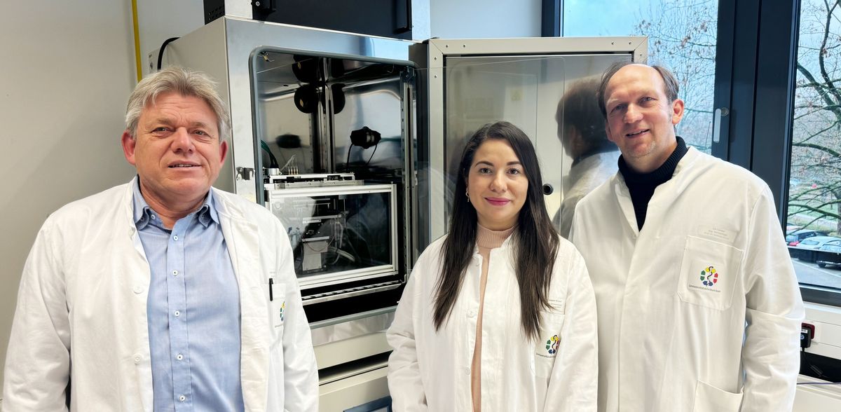
Scientists from the Faculty of Medicine, the Department of Electrical Engineering and Information Technology at UDE and the ISAS in Dortmund worked closely together to develop the ComplexEye. "The challenge was to build miniaturized microscopes, make them movable and assemble them so tightly into a system that they can record videos from each of the 384 chambers of a well plate, a common examination cassette," says Dr. Reinhard Viga from the UDE's Department of Electronic Components and Circuits. The electrical engineer led the technical construction of the new microscope. Like the compound eye of a fly, the ComplexEye moves under the well plate and takes pictures simultaneously with all lenses at intervals of eight seconds. The researchers then combine these images to create a time-lapse video. The researchers then use AI to track the migrating cells visible in these videos. In the future, the ComplexEye will be expanded to include additional lenses so that even more images can be captured.
Note: This article has been translated using a computer system without human intervention. LUMITOS offers these automatic translations to present a wider range of current news. Since this article has been translated with automatic translation, it is possible that it contains errors in vocabulary, syntax or grammar. The original article in German can be found here.
Original publication

Other news from the department science


