New insights into lung tissue in Covid-19 disease
Researchers develop new three-dimensional imaging technique to visualize tissue damage in severe Covid-19
Physicists at the University of Göttingen, together with pathologists and lung specialists at the Medical University of Hannover, have developed a three-dimensional imaging technique that enables high resolution and three-dimensional representation of damaged lung tissue following severe Covid-19. Using a special X-ray microscopy technique, they were able to image changes caused by the coronavirus in the structure of alveoli (the tiny air sacs in the lung) and the vasculature.
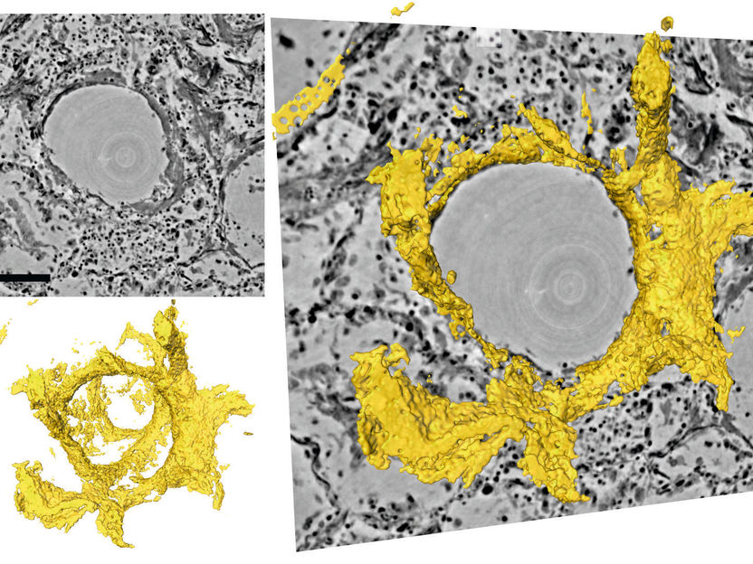
Sections through the three-dimensional reconstruction volume (upper left, grey) around a pulmonary alveolus with hyaline membrane (lower left, yellow). On the right, the images are superimposed. In the centre is the air bubble (alveolus). The electron density is represented by different shades of grey. On the inside of the air bubble is a layer of proteins and dead cell residues, the "hyaline membrane". This deposit, which can be represented in its three-dimensional structure for the first time by the new method, reduces the gas exchange and leads to respiratory distress.
Tim Salditt, Marina Eckermann
In severe Covid-19 disease, the researchers observed significant changes in the vasculature, inflammation, blood clots and “hyaline membranes”, which are composed of proteins and dead cells deposited on the alveolar walls, which make gas exchange difficult or impossible. With their new imaging approach, these changes can be visualized for the first time in larger tissue volumes, without cutting and staining or damaging the tissue as in conventional histology. It is particularly well suited for tracing small blood vessels and their branches in three dimensions, localizing cells of the immune systems which are recruited to the inflammation sites, and measuring the thickness of the alveolar walls. Due to the three-dimensional reconstruction, the data could also be used to simulate gas exchange.
“Using zoom tomography, large areas of lung tissue embedded in wax can be scanned enabling detailed examination to locate particularly interesting areas around inflammation, blood vessels or bronchial tubes,” says lead author Professor Tim Salditt from the Institute of X-ray Physics at the University of Göttingen. Since X-rays penetrate deep into tissue, this enables scientists to understand the relation between the microscopic tissue structure and the larger functional architecture of an organ. This is important, for example, to visualize the tree of blood vessels down to the smallest capillaries.
The authors foresee that this new X-ray technique will be an extension to traditional histology and histopathology, areas of study which go back to the 19th century when optical microscopes had just become available and pathologists could thereby unravel the microscopic origins of many diseases. Even today, pathologists still follow the same basic steps to prepare and investigate tissue: chemical fixation, slicing, staining and microscopy. This traditional approach, however, is not sufficient if three-dimensional images are required or if large volumes have to be screened, digitalized or analysed with computer programmes.
Three-dimensional imaging is well known from medical computerized tomography (CT). However, the resolution and contrast of this conventional technique are not sufficient to detect the tissue structure with cellular or sub-cellular resolution. Therefore, the authors used “phase contrast”, which exploits the different propagation velocities of X-rays in tissue to generate an intensity pattern on the detector. Salditt and his research group at the Institute for X-ray Physics developed special illumination optics and algorithms to reconstruct sharp images from these patterns, an approach which they have now adapted for the study of lung tissue affected by severe progression of Covid-19. The Göttingen team could record lung tissue at scalable size and resolution, yielding both larger overviews and close-up reconstructions. Depending on the setting, their method can even yield structural details below the resolution of conventional light microscopy. To achieve this, the researchers used highly powerful X-ray radiation generated at the PETRAIII storage ring of the German Electron Synchrotron (DESY) in Hamburg.
As was the case when the modern microscope was invented 150 years ago, significant progress has resulted from collaboration between physicists and medical researchers. The interdisciplinary research team hopes that the new method will support the development of treatment methods, medicines to prevent or alleviate severe lung damage in Covid-19, or to promote regeneration and recovery. "It is only when we can clearly see and understand what is really going on, that we can develop targeted interventions and drugs," adds Danny Jonigk (Medical University Hannover), who led the medical part of the interdisciplinary study.
Original publication
Other news from the department science


Most read news


More news from our other portals


Last viewed contents
Enantiomorph distribution maps for metals and metallic alloys
Researchers find metal gets stronger with three or more line dislocations
General Lab and Labco to create a leading European medical diagnostics Group
A new iconic drug information system inspired by road signs
The Science Advisory Board report shows increase in usage of key protein research techniques

Science on the back burner: European laboratories struggle with massive restrictions - Great sustainability ambitions and willingness to innovate despite the need to save money
Genzyme Launches Key Test To Monitor Gleevec Resistance - Two Additional Personalized Medicine Tests Also Available
Michael Siekiera joins management board at Hahnemühle
Measurement of lower aldehydes especially acrolein with the 2-HMP method - GC method
BIOTECON Diagnostics signs license agreement with Applera Corporation