In a class of its own - virtually noise-free images!
The new "cooled cameras" with controlled double stage Peltier cooling provide unbeatable performance in all areas. They offer outstanding performance for low light applications with extremely long exposure times, e.g. in the area of fluorescence microscopy, astronomy, electrophoresis and gel documentation. The cooled cameras feature a high-quality 2/3"CCD sensor that stands out with its extremely high sensitivity, a high resolution and excellent quantum efficiency. The low-noise cameras work with 12-bit pixel depth. The interface can be chosen between Fire Wire, Camera Link (also PCMCIA) or Gigabit Ethernet. For virtually noise-free images: At room temperature the sensor is cooled to -15°C with the controlled double stage cooling (unregulated max. T = -42°). The enormous reduction of thermic noise guarantees an almost noise-free signal (66 dB SNR) even for exposure times of several minutes. The electronic noise is minimized due to the use of high-quality components and intelligent circuitry. Furthermore, the readout noise can be reduced using the Slow Scan Mode with a lower clock rate. The "signature cameras"are also available as cooled versions.
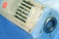
new "Cooled Cameras"
Kappa opto-electronics GmbH
Other news from the department research and development


Most read news


More news from our other portals


See the theme worlds for related content
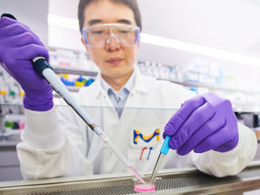
Topic world Fluorescence microscopy
Fluorescence microscopy has revolutionized life sciences, biotechnology and pharmaceuticals. With its ability to visualize specific molecules and structures in cells and tissues through fluorescent markers, it offers unique insights at the molecular and cellular level. With its high sensitivity and resolution, fluorescence microscopy facilitates the understanding of complex biological processes and drives innovation in therapy and diagnostics.