PET detects neuroinflammation in multiple sclerosis
The triggers of autoimmune inflammation in multiple sclerosis (MS) have eluded scientists for many years, but molecular imaging is bringing researchers closer to identifying them, while providing a means of evaluating next-generation therapies for MS.
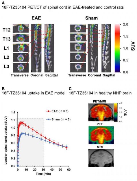
A. Increased F-18 TZ3504 uptake was observed in the inflamed lumbar spinal cord of EAE-treated animals in the rat model of MS compared to sham control rats. Representative sagittal, coronal and transverse views of the thoracic and lumbar spine are shown. B. The time activity curve of F-18 TZ3504 uptake in the lumbar spinal cord was significantly higher for the first 30 minutes in EAE-treated rats. C. F-18 TZ3504 was able to cross the blood brain barrier and showed homogeneous distribution in the brain of a healthy nonhuman primate.
Mallinckrodt Institute of Radiology at the Washington University School of Medicine, St. Louis, Mo.
More than 2.3 million people are affected by MS worldwide, according to estimates from the National Multiple Sclerosis Society. MS is marked by inflammation and the systematic destruction of neuronal fibers, specifically myelin, in the nervous system. Myelin is the fatty layer that both protects the fibers and increases the speed of signaling along the axon of nerve cells. Similar inflammatory processes are typical in the pathology of other neurodegenerative diseases such as Parkinson's and Alzheimer's, gastrointestinal diseases like Crohn's and ulcerative colitis, and the vascular inflammation that leads to atherosclerosis.
"Inflammation is the body's physiological defense to harmful stimuli and it plays a critical role in the immune response to injury and infection," said senior investigator, Zhude Tu, PhD, professor of PET radiochemistry at Washington University School of Medicine in St. Louis, Mo. "However, despite the benefits of acute inflammation in promoting healing, these same processes are associated with numerous pathological conditions when chronic inflammation is left unchecked."
This study furthers a growing body of research pointing to a process called sphingolipid signaling as a primary mechanism in inflammatory disease processes. The FDA approval in 2010 of fingolimod for relapsing MS further supports the hypotheses that the sphingosine-1-phosphate receptor 1 (S1P1) is an ideal biomarker for imaging and new therapies. Fingolimod works by turning down the autoimmune response via immune cell S1P1.
First author of the study Adam J. Rosenberg, PhD, and his colleagues produced a library of S1P1-targeted small molecules and radiolabeled them with fluorine-18. These radiotracers bind directly to S1P1 receptors and can be imaged with preclinical positron emission tomography (PET), through noninvasive methodology to investigate the physiological functions of S1P1 receptors in animal models as a precursor for human studies. In this case, researchers imaged S1P1 in rodent models of inflammatory disease and healthy controls. They found that the PET imaging agents not only were able to detect an increase in S1P1 expression in animals with an inflammatory response when compared to healthy controls, but that the compounds also crossed the blood brain barrier in healthy animals, a significant limiting factor in the development of central nervous system drugs.
"These compounds represent promising PET tracers for imaging MS and other inflammatory diseases by quantitative assessment of S1P1 expression in the body," said Tu.
Other news from the department science


