Electron microscopy: Nano-reporter proteins make invisible processes visible
Genetically encoded nano-barcodes
How do the nerve cells in our brain communicate with each other? What processes take place when T cells render cancer cells harmless? Details of the mechanisms at the cellular level remain hidden from view. Now, special reporter proteins developed by a research team led by the Technical University of Munich (TUM) may help unveil these mechanisms.
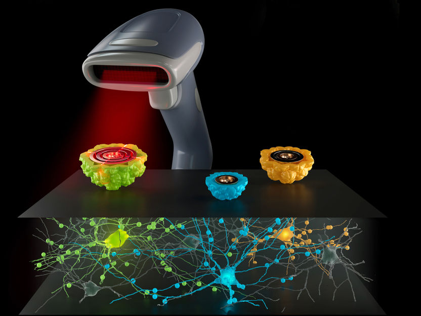
A team around Prof. Gil Gregor Westmeyer has developed a new barcoded gene reporter system for electron microscopy.
Barth van Rossum
Peering through an electron microscope provides scientists the deepest view into cellular structures – the resolution lies in the sub-nanometer range. Even cell components like mitochondria or connections between nerve cells can be discerned. Nonetheless, many important structures and processes remain invisible. "This is somewhat like looking at a city map," explains Gil Gregor Westmeyer, Professor of Neurobiological Engineering at TUM and Director of the Institute for Synthetic Biomedicine at Helmholtz Munich. "It is sufficient to get a visual impression of the surroundings and see where the roads are. But it doesn't tell us how often traffic lights are switched, how much traffic there is at any given point, and when or where something is currently under reconstruction."
But the ability to intervene in faulty processes, or to recreate them in artificial tissues and organs, absolutely requires an understanding of the processes within and between cells. Westmeyer and his colleagues have thus developed a so-called genetic reporter system that does the reconnaissance work within the cells for them. The gene reporters are protein capsules just large enough to be resolved by an electron microscope.

Identification by barcodes
The capsules are produced by the cells themselves. Their genetic blueprints are attached to specific target genes. The reporter proteins are produced when the target genes become active. The basic principle behind this method is already a standard procedure in light microscopy. There, researchers work with fluorescent proteins. However, this method is not suitable for electron microscopy, because rather than colors, different shapes are distinguished based on their electron densities, for example.
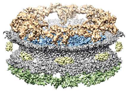
The researchers exploited this by incorporating metal-binding proteins into differently-sized capsules. These "EMcapsulins" appear as variously sized concentric circles under the electron microscope and can be quickly identified and assigned like barcodes using artificial intelligence.
Making invisible structures visible
So, how exactly can the researchers utilize these reporter proteins? On the one hand, they can use them to indicate the activity of certain genes, but also to discover structures that would otherwise not be visible under an electron microscope – e.g. electrical synapses between nerve cells or receptors that influence the interaction between cancer cells and T cells.
"If we also give the EMcapsulins fluorescent properties, this will allow us to initially examine structures in living tissue using light microscopy," says Felix Sigmund, first author of the study. In the process, striking dynamics and structures could be observed, which in a next step can then be highly resolved under an electron microscope.
"It is also conceivable to in the future deploy the reporter proteins as sensors that change their structure, for example, when a cell becomes active. In this way, the relationships between cell function and cell structure can be better elucidated, which is also pertinent to understanding disease processes, as well as to produce therapeutic cells and tissues," adds Westmeyer.
To this end, the researchers will also use the new Electron Microscopy Facility at TUM and collaborate with the new TUM Center for Organoid Systems (COS).
Original publication
F. Sigmund, O. Berezin, S. Beliakova, B. Magerl, M. Drawitsch, A. Piovesan, F. Gonçalves, S.-V. Bodea, S. Winkler, Z. Bousraou, M. Grosshauser, E. Samara, J. Pujol-Martí, S. Schädler, C. So, S. Irsen, A. Walch, F. Kofler, M. Piraud, J. Kornfeld, K. Briggman, G. G. Westmeyer: Genetically encoded barcodes for correlative volume electron microscopy, Nature Biotechnology (2023)
Augmenting electron microscopy with barcoded gene reporters, Nature Biotechnology Research Briefing (2023)
Other news from the department science


