Small step towards growing tissue in the lab
University of Adelaide mathematicians have devised a method for identifying how cell clusters have formed by analysing an image of the cluster.
Published in the Journal of Theoretical Biology, their mathematical modelling tool will be useful in helping biologists and tissue engineers to move towards growing human tissue such as liver in the laboratory.
"When any tissue or organ develops, the cells have to organise themselves into the correct structure," says Dr Edward Green, researcher in the University's School of Mathematical Sciences. "This self-organisation process is important in regenerative medicine where scientists are trying to grow tissues in the laboratory. Getting the right structure is key to ensuring the tissue is viable and functional.
"We know that the control of the organisation process is very complex, and it's still not well understood, which is why we're using modelling to explore simple examples like cluster formation. We looked at two main ways of producing cell clusters – by attraction through chemical and other signals and by proliferation (cells dividing).
"The idea behind our research is that, for any particular cell type, if you are trying to get cells to organise in certain ways, you need to know how they are behaving. We show how you might be able to analyse this using a combination of models and image analysis."
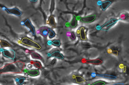
The paper introduces a quantitative measure of the pattern of clustering from an image, producing a statistic called the 'pair correlation function' which shows the relationship between cells.
"The two clustering mechanisms produce different patterns. In some cases you can spot the differences simply by looking, but the pair correlation function allows you to distinguish them, even when you can't see any obvious differences between the pictures by eye," says Dr Green.
They validated their mathematical model experimentally using cells with known clustering mechanisms in collaboration with Queensland University of Technology.
"Our tool gives a basic understanding of the process in clustering," says co-author Dr Ben Binder, Senior Lecturer in the School of Mathematical Sciences. "It will be useful in assessing what factors may be used to enhance the process of growing cells.
"Next steps will be feeding experimental data back into the model to simulate biological processes. Instead of running lengthy and expensive experiments, we can look at the potential effects of different factors through the computer."
Other news from the department science