New technology allows scientists to watch cancer cells in action at unprecedented resolution
Affinity capture devices provide a platform for viewing cancer cells and other macromolecules in dynamic, life-sustaining liquid environments
A photograph of a polar bear in captivity, no matter how sharp the resolution, can never reveal as much about behavior as footage of that polar bear in its natural habitat. The behavior of cells and molecules can prove even more elusive. Limitations in biomedical imaging technologies have hampered attempts to understand cellular and molecular behavior, with biologists trying to envision dynamic processes through static snapshots.

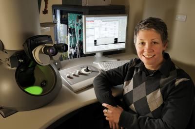
Deborah Kelly, an assistant professor in the Virginia Tech Carilion Research Institute, has now developed a novel technology platform to peer closely into the world of cells and molecules within a native, liquid environment.
Virginia Tech Photo
Deborah Kelly, an assistant professor in the Virginia Tech Carilion Research Institute, has now developed a novel technology platform to peer closely into the world of cells and molecules within a native, liquid environment.
Kelly and colleagues have developed a way to isolate biological specimens in a flowing, liquid environment while enclosing those specimens in the high-vacuum system of a transmission electron microscope (TEM). The TEM liquid-flow holder, developed by Protochips Inc. of Raleigh, N.C., accommodates biological samples between two semiconductor microchips that are tightly sealed together. These chips form a microfluidic device smaller than a Tic Tac. This device, positioned at the tip of an EM specimen holder, permits liquid flow in and out of the holder. When these chips are coated with a special affinity biofilm that Kelly developed, they have the ability to capture cells and molecules rapidly and with high specificity. This system allows researchers to watch - at unprecedented resolution - biological processes as they occur, such as the interaction of a molecule with a receptor on a cell that triggers normal development or cancer.
"With this new technology, we can capture and view the native architecture of cells and their surface protein receptors while learning about their dynamic interactions, such as what happens when cells interact with pathogens or drugs," said Kelly. "We can now isolate cancer cells, for example, and view the early events of chemotherapy in action."
Kelly had previously worked with colleagues at Harvard Medical School to develop a way to capture protein machinery in a frozen environment. "But life moves," said Kelly. "It's better if biological processes don't have to be paused or frozen in order to be studied, but can be viewed in dynamic and life-sustaining liquid environments."
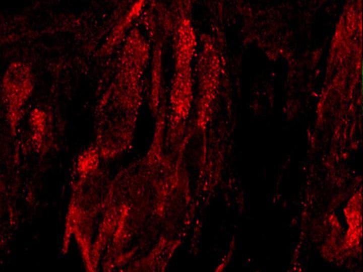
Kelly's affinity capture device, in combination with high-resolution TEM, helps bridge the gap between cellular and molecular imaging, allowing researchers to achieve spatial resolution as high as two nanometers. "This device allows us to see new features on the surface of live cancer cells, providing new targets for drug therapy," Kelly said. "With this resolution, scientists may even be able to visualize disease processes as they unfold."
Original publication
Katherine Degen, Madeline Dukes, ustin Tanner, Deborah Kelly; "The development of affinity capture devices - a nanoscale purification platform for biological in situ transmission electron microscopy."; RSC Advances 2012.
Most read news
Original publication
Katherine Degen, Madeline Dukes, ustin Tanner, Deborah Kelly; "The development of affinity capture devices - a nanoscale purification platform for biological in situ transmission electron microscopy."; RSC Advances 2012.
Organizations
Other news from the department science

Get the analytics and lab tech industry in your inbox
By submitting this form you agree that LUMITOS AG will send you the newsletter(s) selected above by email. Your data will not be passed on to third parties. Your data will be stored and processed in accordance with our data protection regulations. LUMITOS may contact you by email for the purpose of advertising or market and opinion surveys. You can revoke your consent at any time without giving reasons to LUMITOS AG, Ernst-Augustin-Str. 2, 12489 Berlin, Germany or by e-mail at revoke@lumitos.com with effect for the future. In addition, each email contains a link to unsubscribe from the corresponding newsletter.
Most read news
More news from our other portals
Last viewed contents

How do we taste sugar, bacon and coffee? - A new chemical pathway that helps the brain detect sweet, savory and bitter flavors

The smallest test tube in the world - Prof. Dr. Helmut Schwarz receives the "BBVA Foundation Frontiers of Knowledge Award"
FRITSCH Milling and Sizing subsidiary officially dedicated in Beijing

New technique for imaging cells and tissues under the skin developed
PLIVA Opens New Biostudy Center in Goa
Development Cooperation for New Molecular Diagnostic Assays between Bruker Daltonik, PANATecs and the University of Goettingen


