Tomoscopy: new imaging method
Thanks to a world record in tomography, synchrotron radiation can be used to watch how metal foam forms
An international research team at the Swiss Light Source (SLS) has set a new tomography world record using a rotary sample table developed at the HZB. With 208 three-dimensional tomographic X-ray images per second, they were able to document the dynamic processes involved in the foaming of liquid aluminium.
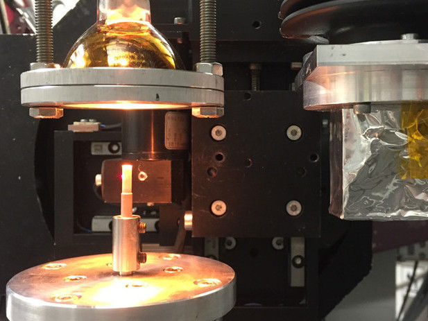
The precision rotary sample table designed at the HZB turns around its axis at several hundred revolutions per second with extreme precision.
© HZB
The precision rotary sample table designed at the HZB rotates around its axis at several hundred revolutions per second with extreme precision. The HZB team headed Dr. Francisco García-Moreno combined the rotary sample table with precision optics and achieved a world record of over 25 tomographic images per second using the BESSY II EDDI beamline in 2018.
Now the team, together with the group headed by Prof. Marco Stampanoni from the Paul Scherrer Institute (PSI), has achieved a new world record at SLS. To accomplish this, they set up the rotary sample table together with improved optics, and used ultra-fast image acquisition & data transfer rates at the SLS's TOMCAT instrument. “Over 200 tomographic images per second can now be acquired – and that during measurement intervals of only several minutes”, says García-Moreno. The term tomoscopy was coined for this new imaging method.
Tomoscopy: new imaging method
Dr. Christian Schlepütz of PSI emphasises: “The enormous volume of data packets generated during each tomoscopy has to be transmitted and stored at the extremely high data rate of eight gigabytes per second."
Each individual image must be calculated from the measurement data. The images then receive additional automatic processing that facilitates quantitative analysis. In order to handle the processing of several terabytes of data from each experiment, Dr. Paul Kamm from the HZB has developed and implemented unique dedicated processing software.
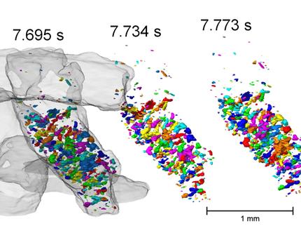
The partners in this collaboration have used the new imaging method to observe dynamic processes in great detail at high temporal resolution that occur during the foaming of liquid aluminium. In this way, processes taking place during the formation of foam in molten metals can be investigated and better understood. This is important in order to achieve optimum material distribution and uniform pore formation in the foam, which is later cured, so that the foam can be used in lightweight construction.
Metal foams for lightweight construction
Metal foams are an important class of materials for lightweight construction, and they are an advantageous subject of investigation for the newly developed imaging method, since liquid metal is largely insensitive to radiation damage, and the imaging speeds achieved are extremely well-suited to foaming phenomena.
Ultrafast computer tomoscopy could also provide interesting insights into many other processes. For example, it could be used to investigate how materials change during laser welding or what happens when batteries overheat due to short circuits (thermal runaway).
The researchers at the HZB and PSI are now working on increasing the rotational speed in order to further increase the temporal resolution of the measurements.
Original publication
"Using X-ray tomoscopy to explore the dynamics of foaming metal"; Francisco García-Moreno, Paul Hans Kamm, Tillmann Robert Neu, Felix Bülk, Rajmund Mokso, Christian Matthias Schlepütz, Marco Stampanoni, John Banhart; Nature communications; 2019
Other news from the department science


