X-ray View into Cerebellum
Researchers at the University and University Hospital of Basel succeed in imaging microscopically small structures of the human brain in three dimensions and automatically detecting the number of Purkinje cells in this tissue.
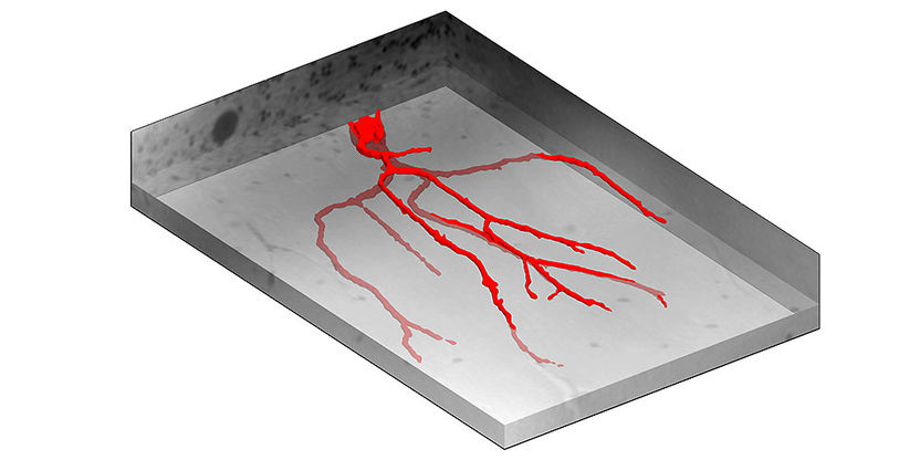
Surface representation of a Purkinje cell with the main part of its dendritic tree, from a microscopic part of the three-dimensional data. The volume corresponds to about 300 x 250 x 50 micrometre and consists of 56 million voxels.
University of Basel, Department of Biomedical Engineering
The human being consists of thousand times more cells than the Milky Way has stars. Therefore, it is a remarkable challenge to three-dimensionally visualize even a part of the human brain on the cellular level.
By the application of a novel X-ray imaging technique and the development of a specific mathematical filter, Dr. Simone Hieber and Prof. Bert Müller of the Department of Biomedical Engineering at the University of Basel succeeded in visualizing such structures and automatically identifying certain neuronal cells in the cerebellum, namely the Purkinje cells.

Imaging using phase contrast
In this tomography study the researchers scanned a specimen of brain tissue, about 40 cubic millimeters in size, using synchrotron radiation to determine the local phase shifts. This approach provides better contrast than conventional X-ray techniques that rely on the attenuation of X-rays. The pixel size was set to 1.75 and 0.45 micrometers, which is a hundred times smaller than the diameter of a human hair.
The obtained three-dimensional data set shows not only the individual Purkinje cells, but also clearly shows their dendritic trees and their nucleoli inside the nucleus, i.e. sub-cellular structures.
Automatic detection of cells
Since the manual analysis of such huge data sets is barely accomplishable, the automatic identification of the Purkinje cells was realized by an in-house developed segmentation algorithm that recognized cells according to their characteristic anatomy. Thus, the researcher could identify thousands of Purkinje cells and determine their surface density with unrivaled accuracy. This allows for the estimation of age or health stage of the human after death.
The validation of the results was performed using histological slides prepared subsequent to the non-destructive tomography.
High resolution imaging of tissue structures
A detailed insight into the cellular structures of the cerebellum enables, for example, a better description of motor function, coordination, and balance regulation. Moreover, morphological changes due to disease such as neurodegeneration should become better recognized on the basis of the three-dimensional imaging data. In combination with the specific software this approach could contribute to a better understanding and treatment of neurodegenerative diseases.
Original publication
Other news from the department science


