How artificial intelligence can improve protein detection
Using AI to move the boundaries of microscopy
Small proteins play a critical role in the regulation of immune response, inflammation and neurodegenerative diseases. In order to better detect and study them, scientists at the Max-Planck-Institute for the Science of Light and FAU have combined one of the most effective microscopy methods, called iSCAT, with artificial intelligence.
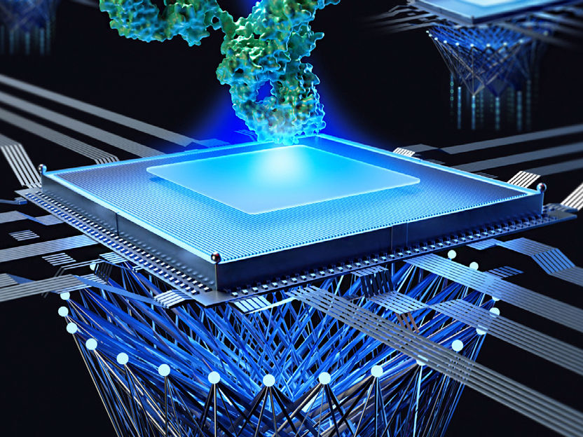
Through a combination of highly advanced microscope technology in iSCAT and multiple machine learning techniques, the team from MPL and FAU continue to push the boundaries of optical sensing.
Mahyar Dahmardeh
Biological molecules such as proteins are central constituents of all living systems and dictate all physiological reactions in health and disease conditions. In particular, many small proteins play critical roles in the regulation of immune response, inflammation, and neurodegenerative disease. Fast and non-invasive protein detection methods can therefore help us create improvements in the areas of disease diagnosis and drug development.
Traditional protein detection methods involve labeling the protein with a fluorescent or radioactive tag, in order to track and detect them. However, these methods have been proven to be quite costly, and time-consuming. Even more problematic is the fact that these labels may alter the function of the studied protein, rendering any gathered data unreliable. As scientific interest in the functions of proteins has increased in recent years, so has the interest in label-free detection methods. One such method, which is now widely regarded as one the most effective and sensitive label-free and real-time protein detection techniques, is interferometric scattering microscopy (iSCAT).
iSCAT is based on the sensitive detection of light scattered by individual proteins through interferometry. As individual proteins sediment out of a buffer onto a cover glass, the tiny shadow of the protein cast on a camera gives information about its size and mass. Hence, the method is also known as mass photometry. However, a combination of technical noise sources and speckle-like background fluctuations has previously limited the sensitivity of iSCAT detection to proteins larger than about 40 KDa.
Using AI to move the boundaries of microscopy
In order to push the sensitivity of iSCAT even further, a team from MPL around the managing director Vahid Sandoghdar composed of electrical engineer Mahyar Dahmardeh, computer scientist Houman Mirzaalian and physical chemist Hisham Mazal has collaborated with Harald Köstler from FAU to employ two machine-learning techniques for detecting proteins at only 10 kDa or less.
In a paper published in Nature Methods, they showed how they can use the iForest algorithm in combination with the FastDVDnet technique to achieve this result. Both are so-called unsupervised machine learning techniques, meaning they do not need to first be trained on a labeled dataset. Unsupervised machine learning is highly desirable in microscopy because it allows identifying patterns and relationships in large data sets without knowing the underlying imaging model. This is especially important when the detection limit is at the edge of the noise level and there is a lack of labeled data to train the network.
FastDVDnet is an advanced image-denoising technique that removes noise from microscopy images using deep neural networks. It is optimized for parallel processing, allowing it to process very large data sets in a relatively short amount of time. In this case, the researchers used FastDVDnet to identify iSCAT images of proteins from the recorded video sequences. Spatiotemporal features extracted by FastDVDnet were then used by iForest to cluster the iSCAT data.
The isolation Forest (iForest) unsupervised machine learning algorithm is commonly used for anomaly detection tasks. It is especially well-suited for microscopy because it can handle high-dimensional data with a large number of features, resulting in more accurate and comprehensive results. This is especially useful when analyzing microscopy data, where identifying rare or abnormal features become important. For example, iForest anomaly detection can be used to detect the presence of rare structures within a biological tissue or to identify cells with unusual morphologies. This algorithm can assist in identifying rare or unusual features that traditional analysis methods might very well overlook.
Professor Vahid Sandoghdar reminisces about the hard work by his team, but he is also already looking forward to the next challenge: “We have come a long way since our first report of label-free small protein detection in Nature Communications in 2014. We are determined to push the detection limit further both by improving the physical measurement methods and by developing more sophisticated machine learning algorithms. There is really no fundamental reason why we should not be able to detect molecules below 1kDa, coming close to the weight of even a single lipid molecule.”
Original publication

Other news from the department science


Most read news


More news from our other portals


See the theme worlds for related content
Topic world Protein analytics
Protein analytics provides a deep insight into these complex macromolecules, their structure, function and interactions. It is essential for discovering and developing biopharmaceuticals, understanding disease mechanisms, and identifying therapeutic targets. Techniques such as mass spectrometry, Western blot and immunoassays allow researchers to characterize proteins at the molecular level, determine their concentration and identify possible modifications.