Live-cell microscopy reveals cell migration by direct forces
How do cells move in a certain direction in the body -- go to a wound site and repair it, for example, or hunt down infectious bacteria and kill it?
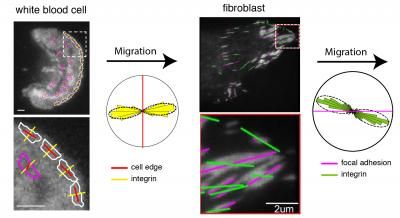
The images show the orientation of integrins (yellow and green lines) at the leading edge (red lines) of a migrating white blood cell and within the focal adhesions (magenta lines) of a migrating fibroblast, analyzed using the instantaneous fluorescence polarization microscope developed at MBL. Circular histograms show that integrins are consistently oriented relative to the direction of migration in both cell types.
(white blood cell) Pontus Nordenfelt, Travis Moore, Shalin Mehta; (fibroblast) Vinay Swaminathan, Shalin Mehta.
Two new studies from the Marine Biological Laboratory (MBL) show how cells respond to internal forces when they orient, gain traction, and migrate in a specific direction. The research, which began as a student project in the MBL Physiology Course and was developed in the MBL Whitman Center, is published in Proceedings of the National Academy of Sciences (PNAS) and this week in Nature Communications.
Both papers focus on the activation of integrins, proteins that allow cells to attach to their external environment and respond to signals coming from other cells. Integrins are transmembrane proteins: part lies on the cell surface and part lies inside the cell. Using a microscope invented at the MBL, the authors showed that when integrins unfurl from the cell surface and bind extracellularly, they simultaneously align in the same direction as a force inside the cell (actin retrograde flow).
"If you think of a cell as a car, the actin flow is the engine," says Clare Waterman, a Whitman Center Scientist from the National Heart, Lung and Blood Institute. "The cell can sit there, idling its engine. But when the integrins activate and bind externally, they are like the tires hitting the road, providing friction. The engine goes into gear and the car moves."
Timothy Springer of Harvard University, who co-discovered the integrin family of proteins in the 1980s and has largely defined their mechanism of activation, and Satyajit Mayor of The National Centre for Biological Sciences, Bangalore, were principal collaborators with Waterman on the project.
The team used a fluorescence polarized light microscope developed by MBL Associate Scientist Tomomi Tani and former Staff Scientist Shalin Mehta (now at Chan Zuckerberg Biohub) to measure -- in real time and with high precision -- the orientation of the integrins on the cell surface.
"It's quite remarkable that you can do that with a microscope," Springer says. "I don't know of any other examples where people have actually measured the orientation of a cell surface molecule."
There are 24 different types of integrins found on human cells. The PNAS paper studies an integrin on fibroblast cells while the Nature Communications paper analyzes an integrin on white blood cells.
"The two integrins we worked on were about as structurally different as you can get in the integrin family," says Springer, yet both types, when activated, oriented in a direction dictated by intracellular actin flow.
"This is really beautiful basic research," Springer says. "While we knew a lot about highly purified integrins in solution, this research gives us specific information about their activation state in living cells."
Waterman was co-directing the MBL Physiology Course when she initiated this research with a group of students, including Vinay Swaminathan and Pontus Nordenfelt. After the course ended, the team added members, including Joseph Mathew Kalappurakkal and Travis I. Moore, and continued to collaborate in the MBL Whitman Center with support from a Lillie Research Innovation Award from the University of Chicago and the MBL.
"The MBL is known for its ability to convene scientific teams with deep interdisciplinary expertise though the communication that flows between its advanced courses, its resident scientists, and the Whitman Center," says David Mark Welch, MBL Director of Research. "In this case, insightful scientists with very different skills -- cell biologists, microscope developers, computational scientists, molecular modelers, protein chemists -- synergized to reveal a fundamentally important driver of cellular migration."
Original publication
Vinay Swaminathanan, Joseph Mathew Kalappurakkalal, Shalin B. Mehta, Pontus Nordenfelt, Travis I. Moore, Nobuyasu Koga, David A. Baker, Rudolf Oldenbourg, Tomomi Tani, Satyajit Mayor, Timothy A. Springer, and Clare M. Waterman; "Actin retrograde flow actively aligns and orients ligand-engaged integrins in focal adhesions"; PNAS; 2017
Pontus Nordenfelt, Travis I. Moore, Shalin B. Mehta, Joseph Mathew Kalappurakkal, Vinay Swaminathan, Nobuyasu Koga, Talley J. Lambert, David Baker, Jennifer C. Waters, Rudolf Oldenbourg, Tomomi Tani, Satyajit Mayor, Clare M. Waterman & Timothy A. Springer; "Direction of actin flow dictates integrin LFA-1 orientation during leukocyte migration"; Nature Comm.; 2017
Other news from the department science


