Novel quantum dots enhance cell imaging
A team of researchers from the University of Illinois at Urbana-Champaign and Mayo Clinic have engineered a new type of molecular probe that can measure and count RNA in cells and tissue without organic dyes. The probe is based on the conventional fluorescence in situ hybridization (FISH) technique, but it relies on compact quantum dots to illuminate molecules and diseased cells rather than fluorescent dyes.
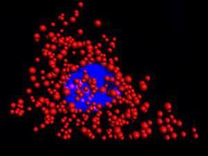
Quantum dots illuminate the locations of individual mRNA as red dots in the cytoplasm of a single HeLa cell. The blue region is the nucleus. This work was a collaborative effort between Illinois Bioengineering and Mayo Clinic researchers.
University of Illinois at Urbana-Champaign Department of Bioengineering
Over the last 50 years, FISH has evolved into a multi-billion-dollar industry because it effectively images and counts DNA and RNA in single cells. However, FISH has its limitations due to the delicate nature of the dyes. For example, the dyes rapidly deteriorate and are not very good at imaging in three dimensions. In addition, conventional FISH can only read out a couple of RNA or DNA sequences at a time.
Quantum dots illuminate the locations of individual mRNA as red dots in the cytoplasm of a single HeLa cell. The blue region is the nucleus. This work was a collaborative effort between Illinois Bioengineering and Mayo Clinic researchers.
Quantum dots illuminate the locations of individual mRNA as red dots in the cytoplasm of a single HeLa cell. The blue region is the nucleus. This work was a collaborative effort between Illinois Bioengineering and Mayo Clinic researchers.
"By replacing dyes with quantum dots, there are no stability issues whatsoever and we can count numerous RNAs with higher fidelity than before," said Andrew Smith, an associate professor of Bioengineering and member of the research team. "Moreover, we uncovered a fundamental limit to the size of a molecular label in cells, revealing new design rules for analysis in cells."
In their latest paper Smith and his team identified an optimal size for quantum dots in order to effectively work with the FISH protocol. This discovery enabled quantum dot-based FISH to match the labeling accuracies currently obtained with organic dyes.
The team created unique quantum dots that are made of a zinc, selenium, cadmium, and mercury alloy and are coated with polymers. "The core of the dot dictates the wavelength of emission, and the shell dictates how much light will be given off," said Smith, who is also affiliated with the Micro + Nanotechnology Lab, Carle Illinois College of Medicine, and Department of Materials Science and Engineering at the University of Illinois.
These dots can emit color independent of the size of the particle, which is not the case for conventional quantum dots. The dots are also small enough (7 nanometers) to fit on a probe that can maneuver between proteins and DNA in a cell, making them more comparable in size to the dyes used in conventional FISH probes.
In experiments with HeLa cells and prostate cancer cells, the researchers found that dye-based FISH cell counts declined rapidly in minutes. The quantum dot-based FISH method provided long-term luminescence to allow counting of RNA for more than 10 minutes, making it possible to acquire 3D cell imaging.
"This is important because images of cells and tissues are acquired slice-by-slice in sequence, so later slices that are labeled with dyes are depleted before they can be imaged," said Smith.
Original publication
Other news from the department science


Most read news


More news from our other portals


See the theme worlds for related content
Topic World Cell Analysis
Cell analyse advanced method allows us to explore and understand cells in their many facets. From single cell analysis to flow cytometry and imaging technology, cell analysis provides us with valuable insights into the structure, function and interaction of cells. Whether in medicine, biological research or pharmacology, cell analysis is revolutionizing our understanding of disease, development and treatment options.