A signal boost for molecular microscopy
Cavity-enhanced Raman-scattering reveals information on structure and properties of carbon nanotubes
carbon nanotubes can be produced with a variety of shapes and properties and are therefore of much interest for widespread applications in fields as diverse as electronics, photonics, nanomechanics, and quantum optics. Hence it is important to have a tool at hand that allows to determine these properties in a quick and precise way. Raman spectroscopy is particularly sensitive for the chemical structure that gives rise to these properties. However, the signals are intrinsically weak and call for enhancement techniques. Now, a team of researchers of the Laser Spectroscopy Division of Prof. Theodor W. Hänsch (Director at the Max Planck Institute of Quantum Optics and Chair of Experimental Physics at the Ludwig-Maximilians-Universität, Munich) has developed a technique, where an optical microcavity is used to enhance Raman scattering signals, and utilized it for molecular diagnostics by combined Raman and absorption imaging. In contrast to other techniques, the new approach only relies on increased vacuum fluctuations of the electromagnetic field inside a cavity, which enables significant enhancement without undesired background, and thereby renders the technique a promising tool for molecular imaging.
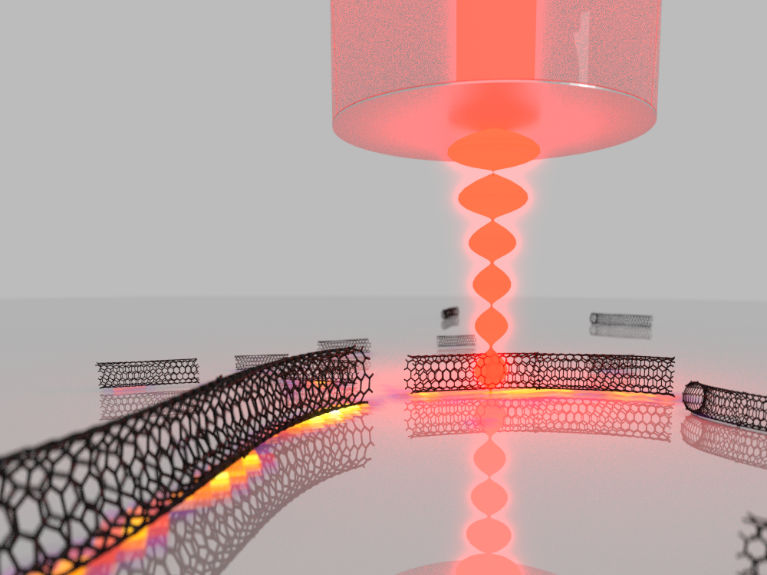
Schematic illustration of the experiment.
Grafik: MPQ, Abteilung Laserspektroskopie
Each molecular species has its own fingerprint of vibrational frequencies which carries information about its chemical structure. Raman spectroscopy allows to optically detect the vibrational spectrum in a powerful manner by inelastic light scattering. As an optical technique, it can enable spatial imaging and thereby combine chemical contrast with high spatial resolution. This capability opens up a large variety of applications for Raman microscopy, ranging from the analysis of biological samples to the characterization of nanomaterials and industrial process monitoring.
In the present study, individual carbon nanotubes are investigated. Nanotubes come along in a variety of diameters and can be either metallic or semiconducting. Raman spectroscopy is particularly sensitive to the molecular structure that governs these properties, and Raman imaging allows to determine this for individual nanotubes. However, conventional Raman scattering suffers from inherently low signal, which is particularly severe for imaging applications and when studying individual nanosystems. “Our approach is to place the sample of nanotubes, dispersed on a substrate, inside of a microscopic cavity, where optical resonances can be harnessed to enhance the Raman scattering process. At the same time, the cavity can be scanned across the sample and focusses the light to a spot size not too far from the diffraction limit, such that high resolution images can be generated”, explains Dr. David Hunger, one of the scientists working on the project. “The cavity amplifies both the Raman scattering process as well as absorption from the sample. This allows one to combine ultrasensitive absorption microscopy with Raman imaging within a single measurement.”
To make the cavity enhancement effect large, ultimately small cavities capable of storing light for many thousands of circulations are required – which is a particular challenge when in addition scanning capabilities for imaging purposes are desired. In the microcavity setup, developed by Dr. David Hunger and his team, one side of the resonator is made of a plane mirror that serves at the same time as a carrier for the sample under investigation. The counterpart is a strongly curved micro mirror on the end facet of an optical fibre. Laser light is coupled into the resonator through this fibre. The plane mirror is moved point by point with respect to the fibre in order to bring the sample step by step into the focus of the cavity mode. At the same time, the distance between both mirrors is adjusted such that the resonance condition for the cavity is matched with a resonance of a Raman scattering process. This requires positioning accuracy in the range of tens of picometers. “To obtain a full Raman spectrum, we step-wise tune the mirror separation to sweep a cavity resonance across the desired spectral range and collect the cavity-enhanced Raman scattering signal,” explains Thomas Hümmer, the leading PhD student at the experiment. “Since the cavity resonances are extremely narrow, this can lead to a spectral resolution way beyond the capabilities of conventional Raman spectrometers.”
At the same time, the Raman signal is strongly enhanced, due to the so-called Purcell effect. This effect comes from the increased vacuum fluctuations and the large photon lifetime inside the microcavity. In the experiment, this leads to an enhancement of the resonant light by up to a factor 320. When comparing the net signal obtained from a single Raman line from the cavity to the signal obtained with the best possible conventional microscope, the cavity experiment achieves a more than 6-fold increase. Further improvements should allow to boost this enhancement by several orders of magnitude in the future.
The full potential of the technique is then demonstrated by cavity-enhanced hyperspectral imaging. In such a measurement, cavity-enhanced Raman spectra are recorded at many locations on the mirror, and a spatial image can be constructed, displaying e.g. the strength or the line shape of Raman lines. “In our experiment we study one particular Raman transition, which is sensitive to the diameter and the electronic properties of the nanotube. From the hyperspectral image we can deduce the size of a large set of individual tubes and determine whether they are metallic or semiconducting,” explains Thomas Hümmer. Such an analysis can provide crucial information about a sample.
The applicability of the method to a large variety of samples makes it a promising tool for single molecule Raman imaging. Furthermore, the scheme could be extended to build Raman lasers with a variety of novel materials, or it might be used to gain quantum control over molecular vibrations.
Original publication

Other news from the department science


Most read news


More news from our other portals


See the theme worlds for related content
Topic World Spectroscopy
Investigation with spectroscopy gives us unique insights into the composition and structure of materials. From UV-Vis spectroscopy to infrared and Raman spectroscopy to fluorescence and atomic absorption spectroscopy, spectroscopy offers us a wide range of analytical techniques to precisely characterize substances. Immerse yourself in the fascinating world of spectroscopy!