New Method Refines Cell Sample Analysis
Innovation in the field of biomedicine
Researchers at the University of Zurich have developed a novel method which increases more than tenfold the number of proteins that can be visualized per sample, making it possible to generate a comprehensive map of cellular organization across the various cellular states. This highly sophisticated and refined view can be used to advance precision medicine and is already being applied in cancer medicine.
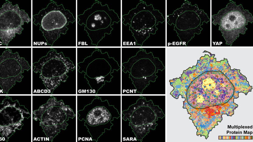
4i is the first imaging technique which gives a multiplexed tissue-to-organelle view of biological samples and links multiplexed information in one and the same experiment.
UZH/Science
Researchers at the University of Zurich have developed a novel method for analyzing cells and their components called Iterative Indirect Immunofluorescence Imaging (4i). This innovation greatly refines the standard immunofluorescence imaging technique used in biomedicine and provides clinicians with an enormous amount of data from each individual sample. 4i makes it possible to observe the spatial distribution of at least 40 proteins and their modifications in the same cell for hundreds of thousands of cells simultaneously at various levels, from the tissue down to the organelle level.
Ten times more proteins visualized at the same time
“4i is the first imaging technique which gives us a multiplexed tissue-to-organelle view of biological samples. We can, for the first time, link multiplexed information derived at the tissue, cellular and subcellular level in one and the same experiment,” says Gabriele Gut, lead author of the study and postdoctoral researcher at the Institute of Molecular Life Sciences at UZH.
Immunofluorescence (IF) uses antibodies to visualize and locate proteins in biological samples. While the standard IF method usually marks three proteins, 4i uses off-the-shelf antibodies and conventional fluorescence microscopes to visualize ten times more proteins by iterative hybridization and removal of antibodies from the sample. “Imagine cell biologists to be journalists. Every experiment is an interview with our cells. With conventional IF I can ask three questions, whereas with 4i I can have a discussion on more than 40 topics,” explains Gabriele Gut.
Map provides systematic survey of cellular landscape
Once acquired, the huge amount of data must then also be able to be analyzed – the next hurdle for the researchers. “We generated images with subcellular resolution for thousands of cells for 40 channels for more than 10 treatment conditions. The human eye and brain cannot process the biological complexity collected by 4i”.
To make full use of the 4i data, Gabriele Gut developed a new computer program for visualization and analysis called Multiplexed Protein Maps. It extracts the multiplexed fluorescence signal for millions of pixels and generates an abstract but representative map of the multiplexed protein distribution in cells.
The researchers were thus able to generate a systematic survey of the cellular landscape: They managed to visualize the spatial intracellular organization of most mammalian organelles along the cell cycle and in different microenvironments.
Advancing precision medicine
The applications for 4i and multiplexed protein maps are manifold, ranging from basic research to precision medicine. “We hope that 4i and multiplexed protein maps will help researchers to understand processes better that have been at the center of biological research for decades,” says Gut. At the same time, the researchers plan to use these technologies to advance the frontiers of precision medicine, particularly in cancer diagnosis and therapy selection.
Original publication
Gabriele Gut, Markus D. Herrmann, Lucas Pelkmans; "Multiplexed protein maps link subcellular organization to cellular states"; Science; 3 August 2018.
Other news from the department science


Most read news


More news from our other portals


See the theme worlds for related content
Topic World Cell Analysis
Cell analyse advanced method allows us to explore and understand cells in their many facets. From single cell analysis to flow cytometry and imaging technology, cell analysis provides us with valuable insights into the structure, function and interaction of cells. Whether in medicine, biological research or pharmacology, cell analysis is revolutionizing our understanding of disease, development and treatment options.