Quantum mechanics rules. It dictates how particles and forces interact, and thus how atoms and molecules work -- for example, what happens when a molecule goes from a higher-energy state to a lower-energy one. But beyond the simplest molecules, the details become very complex. "Quantum mech ... more
Small, beautiful and additive-free
Radically new microscope commercialized
The National Research Council Canada (NRC) recently helped Olympus to design and commercialize a CARS (Coherent Anti-stokes Raman Scattering) microscope.
The new Olympus microscope is based on a femtosecond non-linear optical process called CARS, which stands for Coherent Anti-Stokes Raman Scattering. CARS is a molecule-specific imaging method that permits researchers to see particular chemicals and structures inside living cells, without adding any dyes or stains: CARS is additive-free! While CARS itself is not new, the NRC team developed a simple, robust yet high performance approach that permitted rapid commercialization. CARS microscopy will now be available to biological and medical researchers in a cost-effective system. New opportunities will abound.
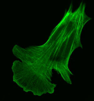
Cells are very small and optical microscopes are required to see them. Unfortunately, the internal components of cells do not come colour-coded for our convenience. To deal with this, scientists have developed, over many decades, various dyes and stains to colour the internal bits of cells so that they could be discerned in an optical microscope. However, if one wants to study processes in live cells busily going about their business, there is a problem. Cellular processes and biochemistry are exceedingly complex and no one knows for sure if these added dyes or stains alter any of these biochemical processes. The best, of course, would be to observe live cells without adding anything at all. Enter CARS microscopy.
CARS is based upon the internal vibrations of molecules. Different molecules have different structures. Due to this, the internal vibrations of these molecules will also be different. CARS recognizes molecules by their "vibrational fingerprint". It is this unique characteristic that allows CARS microscopy to image components inside of cells without adding any dyes or stains.
Compared to Raman microscopy, a well established imaging method that also uses "vibrational fingerprints", CARS is a game changer. For example, a Raman microscopy image that might take 12 hours to scan can now be recorded using CARS microscopy in ½ a second. This allows for real-time movies to be made of live cells and biological tissues.
-
News
Carl Zeiss Licences Digital Pathology from Olympus
Carl Zeiss Microscopy GmbH and Olympus America Inc. have signed a nonexclusive worldwide licensing agreement allowing Carl Zeiss to access an extensive portfolio of patents held by Olympus in the field of digital pathology and virtual microscopy. The patents included in the licensing deal c ... more
Olympus Microscopy has partnered with the Nature Publishing Group to present the Neurotechniques Collection. This compilation of articles will draw together some of the groundbreaking research that has recently been published in Nature Reviews Neuroscience and Nature Methods. Sponsored by O ... more
Olympus Opens Global Research and Development Centre in Munich
Olympus opened a new global Research and Development Centre for Life Science in Munich. Olympus will invest around EUR15m in this project and by 2009 it is expected that 450 people will be employed there. Dr Helmut Köhler, Executive Managing Director of both Olympus Life Science Europa GmbH ... more
-
Products
Spinning Disk Confocal Super Resolution Microscope
High-Level Super Resolution ✓ Suited for Live Specimens ✓ Reveal Super Resolution Details Inside Your Samples more
High-Resolution 3D Images with Low Phototoxicity
Fast high-resolution 3D Imaging ✓ Wide range of samples ✓ Homogeneous illumination with low phototoxicity more
Confocal Laser Scanning Microscope
TruSpectral High-Sensitivity Multichannel Imaging ✓ Macro-to-Micro Imaging and Super Resolution Microscopy ✓ Hybrid Scanning for High-Speed Imaging and Increased Productivity more
- Videos
-
White papers
Benefits of Deep Learning and AI in Microscopy: Label-Free Nucleus Detection in Micro Well Plates
Learn how modern deep learning-based approaches have the potential to reduce or replace fluorescence markers in live cell imaging. Use case included. more
- 1Detect neurodegenerative diseases such as Alzheimer's by a simple eye scan?
- 2Fluorescence microscopy at highest spatial and temporal resolution
- 3The Mechanics of the Immune System
- 4Resolve Biosciences Launches New Era in Single-Cell Spatial Analysis
- 5Quick look under the skin
- 6New ion trap to create the world's most accurate mass spectrometer
- 7How does your computer smell?
- 8Clocking electron movements inside an atom
- 9Sartorius closes 2020 with strong growth
- 10A clear path to better insights into biomolecules