New Method for Analysing Nanoporous Materials
Innovative method opens up new possibilities for materials science
Using only a single electron microscope image, researchers at TU Graz can determine the type and exact position of so-called guest atoms in high-tech materials. They also come closer to solving the mystery of the blue colour of aquamarine.
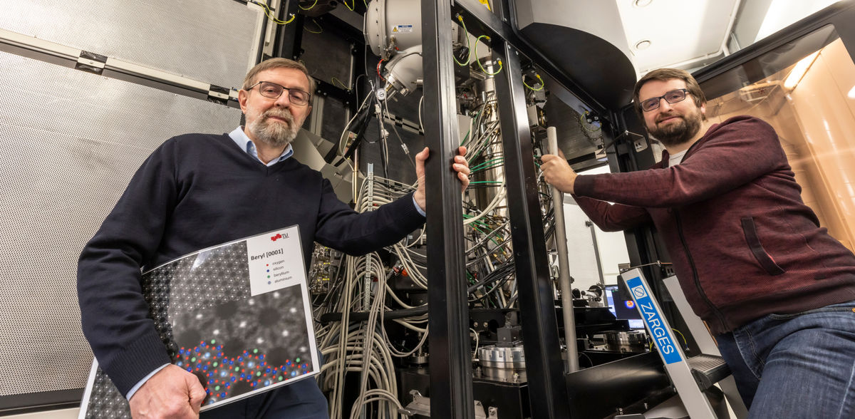
In addition to their main components, the properties of crystalline and nanoporous materials often depend crucially on guest atoms or ions that are embedded in the tiny pores of their lattice structure. This applies to high-tech materials used in sensor or separation technology as well as to natural materials. The bluish gemstone aquamarine, for example, would be colourless without such guest components. Determining the type and position of guest components is difficult, as many materials react sensitively to the radiation emissions from electron microscopes. Thanks to a new method developed by a team led by Daniel Knez and Ferdinand Hofer at the Institute of Electron Microscopy and Nanoanalysis at Graz University of Technology (TU Graz), this can now be done with less radiation and is therefore much easier. “The uniqueness of our method lies in the fact that we can determine the three-dimensional distribution of ions in crystal channels or nanopores based on a single electron microscope image,” says Daniel Knez.
The mysterious blue colour of aquamarine
The researchers developed their method while analysing the gemstone aquamarine. Until now, it was not known where exactly the iron that lends the stone its blue colour is positioned in the crystal. One hypothesis was that individual iron atoms are stuck in the pores and create this effect from there. But this has now been refuted. In their experiments, the researchers have established beyond doubt that there is no iron in the pores, but instead caesium ions. The colour-conferring iron atoms are located in close proximity to the caesium ions, but are integrated into the columns of the crystal lattice.
A single image with atomic resolution as a basis
For their experiments, the researchers recorded a so-called Z-contrast image of the aquamarine crystal at atomic resolution using the ASTEM microscope, a scanning transmission electron microscope. The electron beam of the ASTEM microscope is focused on the surface of the crystal sample, also penetrating into the pores of the material. If it hits ions stored there, they appear as bright dots in the image. Based on the strength of the contrast with empty pores and the neighbouring lattice structures, the researchers can determine the type of embedded ions and also estimate how deep they are located in the pores. These data were statistically analysed and compared with a large number of simulations of the crystal structure in order to be able to estimate the various factors influencing the measured signal. The researchers recently published their findings in the journal Communications Materials.
Innovative method opens up new possibilities for materials science
In addition to basic research, the new method is also suitable for the targeted development of new materials. “Our method can be used to precisely determine the position of doping elements, i.e. targeted function-controlling additives, in nanoporous materials such as zeolites or metal-organic framework compounds,” says Ferdinand Hofer. This facilitates the optimisation of (single-atom) catalysts and solid-state electrolytes in future batteries or the development of biomedical applications for controlling drug uptake.
Original publication
Other news from the department science


Most read news


More news from our other portals


Last viewed contents

A milestone on the pathway to Lab 4.0: A new standard for the smart lab - SPECTARIS presents the first industrial communication standard for laboratory and analytical devices

Fish and seafood - improved trace detection of life-threatening allergy sources - "AQUALLERG-ID": Researchers develop methods for detecting potential food allergens
PerkinElmer appoints President & Chief Operating Officer
ECHA calls for information to avoid unneccessary animal testing
Newly improved NIST reference material targets infant formula analysis

LAUDA appoints Dr. Ralf Hermann as the new General Manager Constant temperature equipment
A relative from the Tianyuan Cave - Ancient DNA has revealed that humans living some 40,000 years ago in the area near Beijing were likely related to many present-day Asians and Native Americans
Measurable for the first time: How bio molecules react to lack of space - Sensor demonstrates lack of space in living cells

Using AI to identify genetic perturbations from cell images - Newly founded start-up aims to use findings to treat previously incurable fibrosis
