Advances in micro-computed tomography
Improved imaging for medicine and material sciences
Researchers in biomedical physics and biology have significantly improved micro-computed tomography, more specifically imaging with phase contrast and high brilliance x-ray radiation. They have developed a new microstructured optical grating and combined it with new analytical algorithms. The new approach makes it possible to depict and analyze the microstructures of samples in greater detail, and to investigate a particularly broad spectrum of samples.
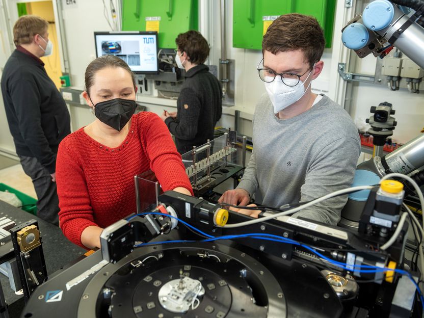
Prof. Dr. Julia Herzen, Professor of Biomedical Imaging Physics at the Technical University of Munich (TUM), has developed a new approach for micro-computed tomography, more specifically for imaging with phase contrast and high brilliance x-ray radiation. The new approach makes it possible to depict and analyze the microstructures of samples in greater detail. Right: Mirko Riedel, one of the two first authors of the publication. Publikation. Back left: Dr. Felix Beckmann, back right: Dr. Julian Moosmann.
René Lahn
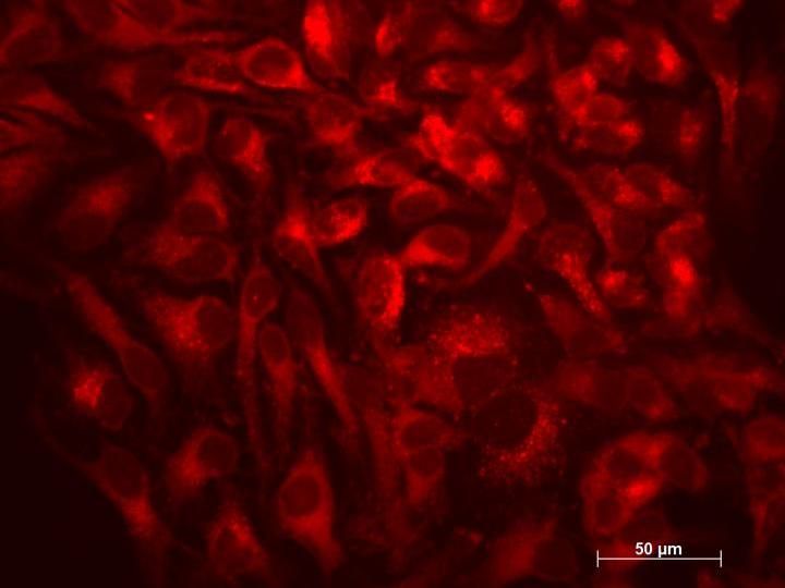
Micro-computed tomography (micro-CT) is an imaging method which generates detailed three-dimensional images of the internal structure of samples with small dimensions. Researchers in biology, medicine or material sciences can use this method to obtain information on the structure and characteristics of tissue and material samples which are important in diagnoses and other analyses.
Micro-CT is based on x-ray images which are reconstructed to form a three-dimensional image. Depending on the sample, different x-ray imaging methods are used in order to achieve the most accurate depiction possible. Here the key parameters are resolution, contrast and the sensitivity of the method used.
X-ray imaging with phase contrast
X-ray imaging with phase contrast is particularly well-suited for investigating soft tissue. The method employs the refraction of the x-rays caused by the sample's structures in order to obtain contrast for these structures and thus to depict soft tissue in greater detail than it is possible with conventional x-ray methods.
In many phase-contrast methods, optical components modulate the x-rays on their way to the detector, resulting in what is referred to as a diffraction pattern at the detector. "When comparing this pattern with and without the sample in the x-ray beam, the refraction of the x-rays on the sample provides information about its characteristics," says Julia Herzen, professor of Biomedical Imaging Physics at the Technical University of Munich (TUM).
Until now inefficient structures such as sandpaper and absorption masks have been used for this type of modulation, but in the meantime a variety of optical gratings are available. "The function of the new optical gratings resembles that of small lenses. The gratings focus the x-rays to form tiny points. This renders the differences in intensity with and without the sample much clearer and makes it possible to visualize even minute differences in the tissue in greater detail," says Prof. Herzen.
High contrast, high resolution and high sensitivity
Physicist Julia Herzen and her team have now introduced a new method for micro-CT with phase contrast using high-brilliance x-ray radiation. The technology is based on a newly developed optical grating referred to as a Talbot Array Illuminator. This new optical element is comparatively easy to produce, is resilient to x-ray radiation and can be used with different energies. This establishes the technically necessary prerequisites for high contrast. The new method enables more efficient use of the radiation dose than with ordinary modulators such as sandpaper and significantly reduces scan times.
"By combining our newly developed Talbot Array Illuminator with new analysis software optimized for the purpose, we've been able to significantly improve imaging and analysis with micro-CT. The new technology is more sensitive than comparable methods in this field. At very high resolutions, it allows to depict soft tissue with higher contrast than previously. High sensitivity is particularly important for example in order to detect fine differences within soft tissue," says Prof. Herzen.
Analysis of a broad spectrum of samples
The new technology can be used to investigate a particularly broad spectrum of samples. Researchers can even simultaneously depict materials of greatly differing compositions, for example water and oil embedded in stone, which was not possible in the past using conventional methods. This provides crucial advantages over conventional methods not only in medicine and biology, but also opens up new application possibilities in material sciences, for example in geology.
Quantitative analysis is possible
"In contrast to previous approaches, our new method also makes quantitative analysis possible. We can make and compare absolute measurements of the electron density of samples, without the need for any assumptions about the samples," explains Prof. Herzen. Further studies will investigate the potential of this new option in a variety of applications.
Original publication
Alex Gustschin, Mirko Riedel, Kirsten Taphorn, Christian Petrich, Wolfgang Gottwald, Wolfgang Noichl, Madleen Busse, Sheila E. Francis, Felix Beckmann, Jörg U. Hammel, Julian Moosmann, Pierre Thibault und Julia Herzen; "High-resolution and sensitivity bi-directional x-ray phase contrast imaging using 2D Talbot array illuminators"; Optica; 8, 1588-1595 (2021).

Other news from the department science


Most read news


More news from our other portals


Last viewed contents
World Finance 100 Award for Carl Zeiss Microscopy - The Carl Zeiss Microscopy business group is one of the world's 100 financially most successful companies

Schizophrenia manifests itself in the brain structure - The symptoms of schizophrenia vary greatly from person to person
The race is on for new lithium ion batteries - Battery consortium headed by BASF receives € 21 million sponsorship from German Ministry of Education and Research
Antibiotic resistance testing no longer impeded by time - Researchers have developed a novel method for antibiotic resistance testing that might transform microbial screening in clinical and research labs
A new way to stimulate cellular recycling process

Thermo Fisher Scientific and QIAGEN N.V. Agree on Amended Terms to Acquisition Agreement - Offer price increased

New insights into the function of a risk factor for Alzheimer’s disease - Researchers decipher the structure-function relationship of the multifunctional protein clusterin for the first time

Rigaku opens Rigaku Technology Center Taiwan - Advancing regional growth through engineering and collaboration in Taiwan and beyond
Multiplexed Morse signals from cells
Lab901 and Biofortuna to collaborate on automated PCR Dx for HLA and Coeliac disease
Closing in on the elusive rotational-vibrational CH5+ spectra
