New method makes masked proteins in nerve cells visible
"This method opens new doors to the study of hard-to-label proteins in living neurons"
The research groups of Prof. Dr. Markus Sauer from the Biocenter and Dr. Gerti Beliu from the Rudolf Virchow Center for Integrative and Translational Bioimaging at the University of Würzburg, as well as the group of Dr. Daniel Choquet from the Bordeaux Neurocampus department of Bordeaux University, developed a new approach that made it possible for the first time to visualize a complex of receptor protein and auxiliary protein in living neurons. "This method opens new doors to the study of hard-to-label proteins in living neurons," Sauer explains.
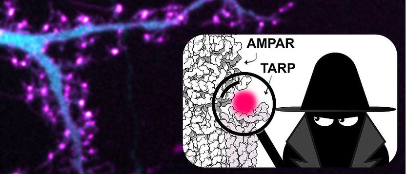
Researchers can now study the interactions of difficult to tag proteins with unrivalled precision. Fluorescence images of living neuron co-expressing a fluorescent protein (cyan) and click-labeled auxiliary protein TARP (magenta).
Choquet group and Beliu group.
Combination of innovative methods and expertise in neuroscience
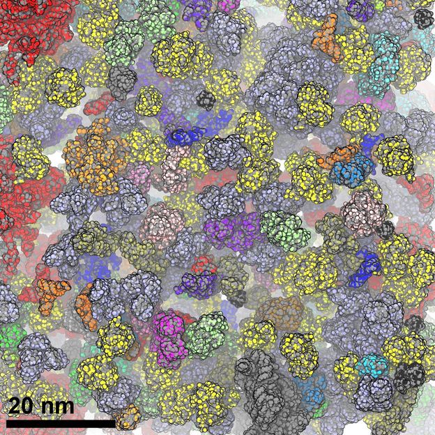
To visualize the proteins, the scientists combined the super-resolution microscopy method dSTORM developed by Sauer with the labeling method of click chemistry. In this technique, an artificial amino acid, the building blocks of which proteins are composed, is incorporated directly into the protein of interest. A tiny dye binds to this artificial amino acid, like a second piece of a puzzle, and thus directly labels the protein. With this new approach, researchers can now see exactly what is happening in a cell, right down to the molecular level.
"In nerve cells, it is particularly important which protein is located where in the cell and with which other proteins it is connected. Because this molecular organization regulates the signal transduction of the neurons and thus such important processes as learning," explains Choquet. Conventional labeling methods, such as labeling with antibodies or linking fluorescent proteins, are limited in their use, particularly in living neurons. These methods can disrupt the normal metabolism of the cell due to the size of the labeling molecules. Therefore, it has also been very difficult to study the exact molecular processes and regulation of the individual proteins in living cells.
Hidden binding site masks receptors
The AMPA receptor is a protein that has attracted great interest in neuroscience. Among other things, the receptor is important for synaptic transmission. Small auxiliary proteins (TARP) bound to the receptor regulate the activity and the localization of it. It was known from structural biology studies that the receptor and auxiliary protein could form a complex. Although the scientists suspected this protein complex must be present at the synapses, it has not yet been possible to label the proteins and visualize them under the microscope.
Now it is clear: the receptors were literally masked. The binding of the auxiliary proteins to the receptors also covered the binding site for the antibody. Since it could not bind, labeling was not possible and the receptors were thus "invisible". With the new method, which was optimized especially for neurons, it is now possible to show the protein complex of receptors and auxiliary protein. The high spatial resolution of dSTORM allows the receptors to be counted individually and their distribution in nerve cells to be studied specifically. The research groups are already working on unmasking other proteins to enable their visualization by super-resolution fluorescence imaging.
Original publication
Diogo Bessa-Neto, Gerti Beliu, Alexander Kuhlemann, Valeria Pecoraro, Sören Doose, Natacha Retailleau, Nicolas Chevrier, David Perrais, Markus Sauer & Daniel Choquet; "Bioorthogonal labeling of transmembrane proteins with non-canonical amino acids unveils masked epitopes in live neurons"; Nature Communications; November 2021

Other news from the department science


Most read news


More news from our other portals


See the theme worlds for related content
Topic world Fluorescence microscopy
Fluorescence microscopy has revolutionized life sciences, biotechnology and pharmaceuticals. With its ability to visualize specific molecules and structures in cells and tissues through fluorescent markers, it offers unique insights at the molecular and cellular level. With its high sensitivity and resolution, fluorescence microscopy facilitates the understanding of complex biological processes and drives innovation in therapy and diagnostics.