A close-up of biological nanomachines
Researchers take a deep look at peroxisomal processes
Every system has its waste disposal system. The cell organelles known as “peroxisomes” dispose toxic substances and fats in the human body, among other things, and, in doing so, they prevent serious illnesses. The “Pex” group of proteins (peroxisomes biogenesis factors) keep these “detox units” functioning properly – and a team of researchers at Münster University headed by Prof. Christos Gatsogiannis have now been the first to show, at the atomic level, how these highly complex processes proceed. The success story – now acclaimed with the study being published in the prestigious journal Nature Communications – was made possible as a result of the University’s new high-tech microscope.
“We can imagine peroxisomes being like miniature factories which specialise in different tasks,” Gatsogiannis explains. “First of all, they are known for ‘detoxifying’ the cell. They act as cellular waste disposal units in our cells.” This waste can be excess fatty acids, for example, or toxic substances from the environment: at least 50 different processes of this kind are attended to by cell organelles only 0.5 micrometres in size (1 micrometre = 1 millionth of a millimetre).
Something that is particularly important for the system is the role played by peroxisomes in fat metabolism. This is because they not only dismantle the fats, they also convert them into usable energy which itself is indispensable for a variety of processes in the body. Without peroxisomes, dangerous quantities of certain fats can accumulate, which would give rise to serious health problems. This is why age-related illnesses are often associated with peroxisomal malfunctions, e.g., loss of hearing or sight, Alzheimer’s, diabetes or cancer.
Each of these processes requires a series of specific enzymes. The peroxisomes, however, are surrounded by a biological membrane which the proteins cannot readily permeate, which means that they have to be imported. This importing mechanism needs energy and a further group of proteins – the Pex group. “Just like a truck, which transports products from one place to another, the transportation of enzymes requires a transportation protein, energy and well-thought out logistics in order to work efficiently,” is the comparison drawn by PhD student Maximilian Rüttermann, a member of the team. “And, again just like a truck, the protein is used again or recycled until ultimately it falls apart or disintegrates.”
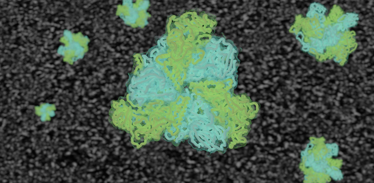
This recycling mechanism is the only energy-intensive step in the entire importing process. The main role is played by the perixisomal AAA-ATPase complex Pex1/Pex6: this “biological nanomachine” unpacks and unfolds the spent proteins so that they can be recycled or disposed of. AAA-ATPases are basically a kind of cellular cleaning crew which keeps the inner surroundings of the cell clean, functional and ready for the demands of life. It is less surprising, therefore, that most of the malfunctions in peroxisomal biogenesis are associated with mutations in Pex1 or Pex6, with up to 60 percent of all cases being attributable to a rare genetic disorder in which the patient’s cells are not able to form peroxisomes. This is something which the general public is not aware of, as patients affected die as a rule just a few days or weeks after their birth – and there is no known cure as yet.
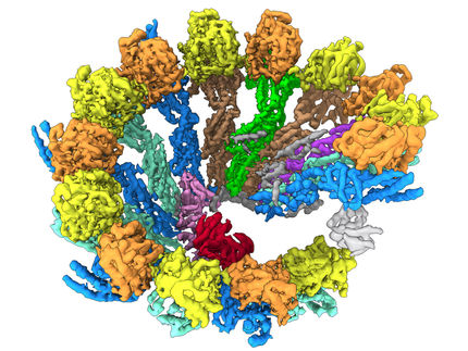
The team of researchers headed by Gatsogiannis has now shown, for the first time and in atomic detail, how the peroxisomal AAA-ATPase processes other enzymes in order to keep the detox units functioning properly. To this end the researchers used the cryogenic electron microscopy method. “Investigating a highly dynamic complex such as AAA-ATPase Pex1 Pex 6 is like watching a car engine running,” says Rüttermann. “You generate millions of images from all angles while it’s running and then, on this basis, produce a three-dimensional model in all its various states.” In spring this year, the team put into operation a state-of-the-art cryogenic electron microscope. The new acquisition, costing 7.5 million euros, makes it possible to investigate proteins and biological nanomachines at the atomic level and thus decrypt the secrets of how cells function.
The high-resolution structures show how the Pex1 and Pex6 proteins work together synchronically. They pull out of the membrane a substrate similar to the import receptors used in order to enable them to be recycled – a unique mechanism, comparable to a row of arms which, step by step, pull a thick rope in pairs and, in the process, untie its knots. “The atomic structures and an understanding of the mechanism of this complex nanomachine now enable us to understand important steps in peroxisome physiology in health and disease,” says Gatsogiannis in conclusion. “It is now possible to relate all known mutations to their function, in order to understand their chemical consequences and, as a result, understand the causes of metabolic disorders.”
Original publication
Other news from the department science


Most read news


More news from our other portals


Last viewed contents
VIAFLO Corporation and INTEGRA Biosciences enter into Strategic Partnership
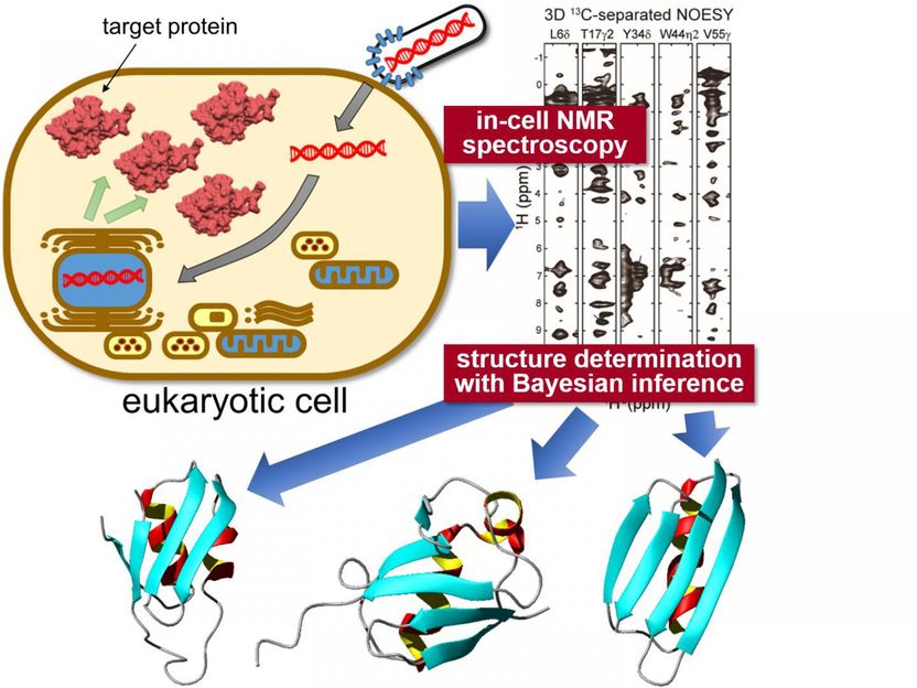
In-situ measurement of 3D protein structure inside living eukaryotic cells - Nuclear magnetic resonance measurement and state-of-the-art computational science reveal protein structures in higher eukaryotic cells
BioTek Instruments Receives Vermont Business Growth Award for Technology