It's not an antibody, it's a frankenbody: A new tool for live-cell imaging
Antibody-based probe works in living systems and targets the classic HA tag
antibodies are the biomolecules our immune systems deploy to find, tag and destroy invading pathogens. They work by binding to specific targets, called epitopes, on the surfaces of antigens - like locks to keys.
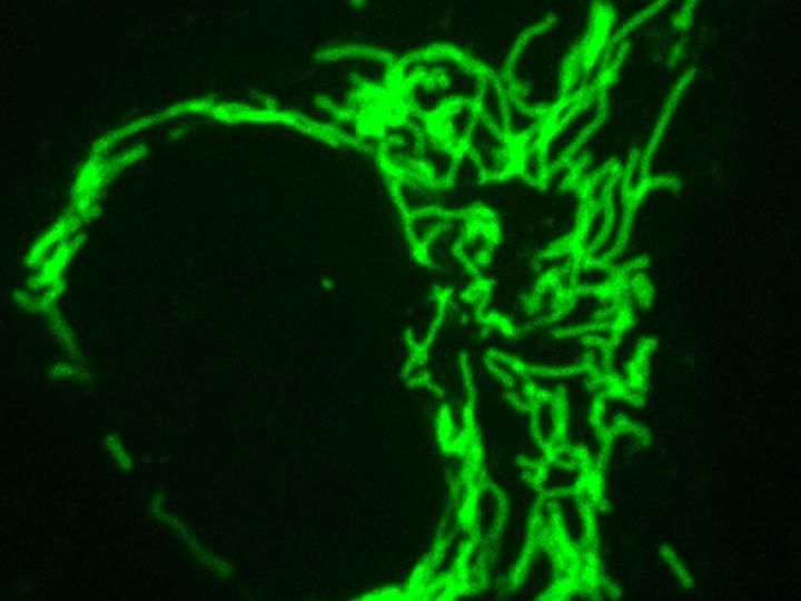
This is a Frankenbody labeling mitochondria.
Ning Zhao/Colorado State University
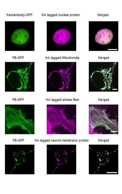
Frankenbodies (green) label nuclear proteins, mitochondria, stress fibers and neuron membranes in living cells.
Ning Zhao/Colorado State University

For many decades, scientists have cleverly exploited this selective tagging mechanism in natural antibodies to engineer antibody-based probes that let them purify and study different types of proteins within cells. One tried and true technique, epitope tagging, involves fusing an epitope to a protein of interest and using fluorescently labeled antibodies to make those proteins visible - but only in fixed, dead cells.
Now, a cross-disciplinary team of researchers from Colorado State University and the Tokyo Institute of Technology have added a new tool to the arsenal of antibody-based probes, but with a powerful distinction: Their genetically encoded probe works in living cells. The work, led by CSU Monfort Professor Tim Stasevich and Tokyo Tech Professor Hiroshi Kimura, is described in Nature Communications.

According to first author Ning Zhao, a postdoctoral researcher in Stasevich's lab who designed most of the experiments, their new antibody-based probe is affectionately called a "frankenbody." Like stitching new limbs on a body, the scientists have taken the binding regions of a normal antibody, the "sticky parts," and grafted them to a different scaffold that remains stable in live cells but retains the specificity of the antibody.
"We're interested in intracellular antibodies because you can use them as imaging reagents in a live cell," said Stasevich, an assistant professor in the Department of Biochemistry and Molecular Biology at CSU. "You don't need a tag, like a Green Fluorescent Protein, because instead you have this fluorescent antibody that will bind to your protein that you want to visualize."
The new probe would be a useful complement to the green fluorescent protein (GFP), a widespread biochemistry tool and subject of a Nobel Prize that involves genetically fusing a light-up green tag to a protein of interest. However, the GFP is limited by its relatively large size and the time it takes to fluoresce; with the CSU researchers' new probe, the tag is smaller and becomes fluorescent faster, so the "birth" of a protein of interest can be captured in real time.
With the goal of making their tool immediately useful, the scientists designed their probe to work with the classic HA tag. HA is a widely used small linear epitope tag that's derived from a portion of the human influenza virus protein hemagglutinin.
"For the longest time, people have been looking at HA-tagged proteins in fixed, dead cells," Stasevich said. "Now we can image the dynamics of those proteins in live cells."
The possibilities of how scientists may use the new probe are limitless. Stasevich's lab is particularly interested in studying RNA translation, and they plan to use their new system to more easily design new RNA imaging experiments.
Added Zhao, the HA tag is tiny - a chain of just nine amino acids - and the probe is genetically encoded on a plasmid that can be easily transferred into a cell. This is in contrast to traditional antibodies, which can cost a lab several hundreds of dollars per order, suffer from lot-to-lot variability, and are difficult to get into cells. The new probe from Stasevich's team therefore provides a low-cost solution for protein and RNA translation imaging.
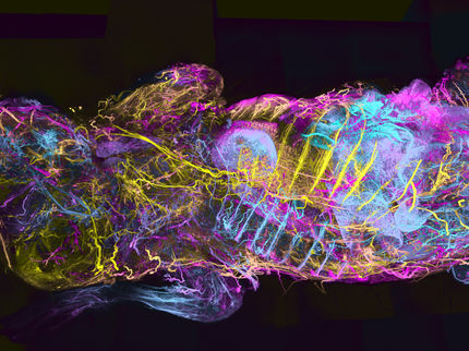
In the paper, the scientists demonstrated some applications, including single-protein tracking, single-RNA translation imaging, and amplified fluorescence imaging in zebrafish embryos. All of these experiments are more challenging when using traditional fluorescent protein tags.
"We have several new imaging reagents in the works that build off of this success, so I see great things ahead," Stasevich said.
Original publication
Other news from the department science


Most read news


More news from our other portals


See the theme worlds for related content
Topic World Cell Analysis
Cell analyse advanced method allows us to explore and understand cells in their many facets. From single cell analysis to flow cytometry and imaging technology, cell analysis provides us with valuable insights into the structure, function and interaction of cells. Whether in medicine, biological research or pharmacology, cell analysis is revolutionizing our understanding of disease, development and treatment options.
Topic world Antibodies
Antibodies are specialized molecules of our immune system that can specifically recognize and neutralize pathogens or foreign substances. Antibody research in biotech and pharma has recognized this natural defense potential and is working intensively to make it therapeutically useful. From monoclonal antibodies used against cancer or autoimmune diseases to antibody-drug conjugates that specifically transport drugs to disease cells - the possibilities are enormous
Last viewed contents
Excellent early results in large OvPlex ovarian cancer biomarker study
Automating Laboratory-On-A-Chip To Cut Healthcare Costs - Computer programming language automates “laboratory-on-a-chip” technologies and has potential to improve and reduce cost of healthcare
