Anchors away – for MRI contrast agents
Italian scientists label cells with a contrast agent and track via MRI, which shows long retention times due to partial internalisation
Silvio Amie and his colleagues from the University of Turin and the University of Eastern Piedmont, in Italy, have anchored a gadolinium (Gd)-based contrast agent on cell surfaces via disulfide bridges.
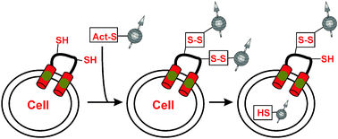
Several kinds of cells show high levels of free exofacial protein thiol (EPT) groups on the extracellular side of the plasma membrane. These are ideal anchorage points for Aime’s Gd-based contrast agent which has a thiol-reactive moiety. Aime has shown that the imaging probe can reversibly bind to the cell surface by the formation of disulfide bridges. Unexpectedly, Aime discovered that the chemical conjugation between the EPTs and the Gd probe triggered an efficient mechanism for the partial internalization of the complex. Consequently, this results in a large amount of the Gd complex being taken up by the cell (not just by the EPTs) and therefore increasing the retention time of the contrast agent within the cell for up to 4 hours.
The visualisation of cell tracking in vivo by imaging techniques is important for the future development of cellular therapies. Aime explains that ‘the development of cell-based therapies will take great advantages from any method allowing for a non-invasive and repetitive monitoring of the delivery of repairing cells to the area of interest. The possibility to visualize cell differentiation or response to biochemical stimuli would be also of immense value.’ Cellular Imaging is a relatively new and very rapidly evolving branch of in vivo imaging techniques devoted to provide answers to these needs. ‘Amongst the available imaging techniques, MRI is one of the most promising, because of its great spatial resolution, lack of invasiveness and ease of adaptability to the clinical practice. Cells to be visualized within a living organism must be suitably labelled for MR detection,’ says Aime.
Aime continues to explain that ‘although a limited number of cell types have been assayed for EPTs levels, we are confident that most cell types do expose appreciable levels of reactive protein thiols.’
Therefore, Aime’s method has potential for becoming a general labelling procedure for all cellular imaging needs or perhaps with future development to become a vehicle for “smart” delivery of contrast agents.
Original publication: Giuseppe Digilio et. al., Chem. Commun., 2009.
Other news from the department science


