Sharp images from the living mouse brain
Max Planck scientists in Goettingen have for the first time made finest details of nerve cells in the brain of a living mouse visible
To explore the most intricate structures of the brain in order to decipher how it functions – Stefan Hell's team of researchers at the Max Planck Institute for Biophysical Chemistry in Göttingen has made a significant step closer to this goal. Using the STED microscopy developed by Hell, the scientists have, for the first time, managed to record detailed live images inside the brain of a living mouse. Captured in the previously impossible resolution of less than 70 nanometers, these images have made the minute structures visible which allow nerve cells to communicate with each other. This application of STED microscopy opens up numerous new possibilities for neuroscientists to decode fundamental processes in the brain.
Every day a huge quantity of information travels not only over our information superhighways; our brain must process an enormous amount of data as well. In order to do this, each of the approximately hundred billion nerve cells establishes contact with thousands of neighboring nerve cells. The entire data exchange takes place via contact sites – the synapses. Only if the nerve cells communicate via such contact sites at the right time and at the right place can the brain master its complex tasks: We play a difficult piece of piano, learn to juggle, or remember the names of people we have not seen for years.
We can learn most about these important contact sites in the brain by observing them at work. When and where do new synapses form and why do they disappear elsewhere? This is not easy to determine, since details in living nerve cells can only be observed with optical microscopes. Due to the diffraction of light, however, structures located closer together than 200 nanometersappear as a single blurred spot. The STED microscopy developed by Stefan Hell and his team at the Max Planck Institute for Biophysical Chemistry is a groundbreaking method devised to surpass this resolution limit. They use a simple trick: Closely-positioned elements are kept dark under a special laser beam so that they emit fluorescence sequentially one after the other, rather than simultaneously, and can therefore be distinguished. Using this technique, Hell's team has been able to increase the resolution by approximately tenfold compared to conventional optical microscopes.
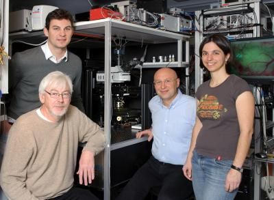
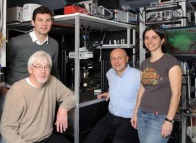
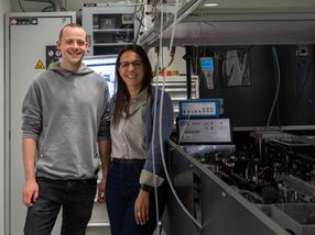
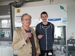
Dr. Heinz Steffens (front left), Dr. Sebastian Berning (back left), professor Dr. Stefan W. Hell (middle), and Dr. Katrin Willig (right) successfully captured high-resolution live images inside a mouse brain.
MPI for Biophysical Chemistry
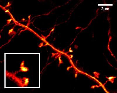
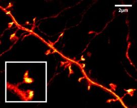
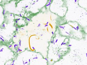
This STED image of a nerve cell in the upper brain layer of a living mouse shows in previously impossible detail the very fine dendritic protrusions of a nerve cell, the so-called spines, at which the synapses are located. The inset shows the mushroom-shaped head of such a dendritic spine at which the nerve cells receive information from their peers.
MPI for Biophysical Chemistry
A vision becomes reality
What was only an ambitious vision a year ago has now become reality: to also study higher living organisms at this sharp resolution in the nanometer range. By looking directly into the brains of living mice using a STED microscope, Hell and his team were the first ones to image nerve cells in the upper brain layer of the rodent with resolution far beyond the diffraction limit.
"With our STED microscope we can clearly see the very fine dendritic structures of nerve cells at which the synapses are located in the brain of a living mouse. At a resolution of 70 nanometers, we easily recognize these so-called dendritic spines with their mushroom- or button-shaped heads," explains Hell. They are the clearest images of these fundamental contact sites in the brain to date. "To make these visible, we take genetically modified mice that produce large quantities of a yellow fluorescing protein in their nerve cells. This protein migrates into all the branches of the nerve cell, even into smallest, finest structures," adds Katrin Willig, a postdoctoral researcher in Hell's department. The genetically modified mice for these experiments originated from the group of Frank Kirchhoff at the Göttingen Max Planck Institute for Experimental Medicine. Images of the nerve cells taken seven to eight minutes apart revealed something surprising: The dendritic spine heads move and change their shape. "In the future, these super-sharp live images could even show how certain proteins are distributed at the contact points," adds Hell. With such increasingly detailed images of structures in the brain, Hell's team hopes to shed light onto the composition and function of the synapses on the molecular level.
Original publication
Sebastian Berning, Katrin I. Willig, Heinz Steffens, Payam Dibaj, Stefan W. Hell: "Nanoscopy in a living mouse brain."; Science 335:551 (2012).
Other news from the department science


Get the analytics and lab tech industry in your inbox
From now on, don't miss a thing: Our newsletter for analytics and lab technology brings you up to date every Tuesday. The latest industry news, product highlights and innovations - compact and easy to understand in your inbox. Researched by us so you don't have to.
Most read news


More news from our other portals


See the theme worlds for related content
Topic world Fluorescence microscopy
Fluorescence microscopy has revolutionized life sciences, biotechnology and pharmaceuticals. With its ability to visualize specific molecules and structures in cells and tissues through fluorescent markers, it offers unique insights at the molecular and cellular level. With its high sensitivity and resolution, fluorescence microscopy facilitates the understanding of complex biological processes and drives innovation in therapy and diagnostics.