The Glowing Brain
Method conveys medical interrelationships quickly and intuitively
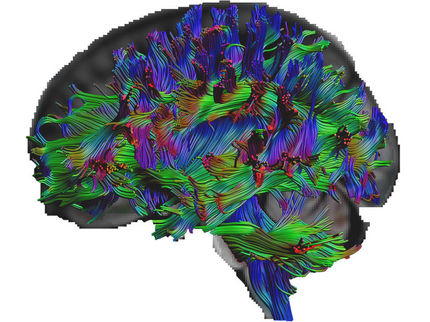
On the monitor, a brain spins slowly and can be examined from every angle. Suddenly, some sections start glowing, first on the side and then the entire back of the head. These spectacular images show which brain areas activate when we speak, see, hear, or touch.. The method originated at the Fraunhofer Institute for Medical Image Computing MEVIS in Bremen. It uses modern visualization technology called ‘physically based rendering’ in combination with medical image data and enriched with clinically relevant supplementary information. On October 1, the method will premiere in an exhibit at the AUDIOVERSUM Science Center in Innsbruck, Austria.
Computer technology now is so powerful that photorealistic animations can even be produced with common PCs and modern graphic cards. Physically based rendering plays an important role here, simulating how individual rays of light spread in specific scenery and how the environment influences each ray in reality. Mirrors reflect these rays; frosted glass weakens and scatters them; obstacles absorb them. As a result, walls, objects, and people appear in naturally pleasing light, wine glasses mirror reflections, objects cast complex shadows. The scene appears as realistic as a photo or a video.
Hollywood directors and computer game programmers regularly use such systems and astound the public with increasingly lifelike computer images. Fraunhofer MEVIS experts use this technique for another purpose: based on medical data from CT or MR images, they produce three-dimensional moving images enriched with supplementary medical information. Their work results in moving images that visualize complex medical relationships in an aesthetical and instructive way. Initial applications include 2D and 3D movies, training materials, and interactive installations. In the long term, the method can be used as an augmented visualization tool in medicine, for example, for diagnosis and operation planning.
Software systems that process medical image data from CT and MR scanners into three-dimensional images have existed for some time. “We took these anatomical images to another level with photorealistic, clinically relevant supplementary information that can be extracted from medical data with the help of special software,” explains Alexander Köhn, software architect at Fraunhofer MEVIS. “This supplementary information fuses with anatomical imagery.”
One example for this ‘Meta-realistic Medical Moving Images’ approach is an interactive image sequence that visualizes the functionality of the human brain. “We want to use the strong visual cognitive ability of the human to transfer complex interrelationships in an intuitive, correct, and fast way,” says Bianka Hofmann, scientific communication specialist at Fraunhofer MEVIS. The researchers first produced high-resolution anatomical 3D images of the brain with an MR scanner. Then, subjects performed different tasks in the scanner, such as reading texts, reciting poems, listening to music, or viewing images. They switched the scanner to a specific mode, allowing it to capture, in high temporal resolution, the effect of blood supply in the brain during the different tasks.
Based on these functional MR images, statistical algorithms determine how intensively the different brain areas participate in the specific tasks. The auditory center on the side of the skull reacts when listening to music and the visual cortex on the back of the head is stimulated when observing images. The higher the intensity, the brighter this particular image volume becomes. When combined with anatomical MR data, images emerge in which the brain areas responsible for seeing or hearing start to glow. With the help of physically based rendering, these images appear extremely realistic.
On October 1, the technique will be unveiled at the AUDIOVERSUM in Innsbruck, Austria during the Long Night of Museums. The Science Center will present an exhibit designed by Fraunhofer MEVIS which allows visitors to discover the brain’s functionality interactively. On September 9 and 10, MEVIS Institute Director Horst Hahn will present sequences from the ‘Meta-realistic Medical Moving Images’ at the Ars Electronica Festival in Linz.
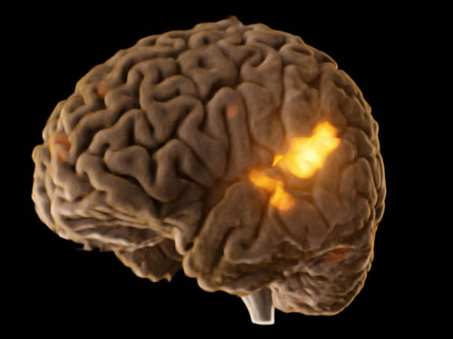
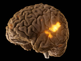
A fusion of anatomical and functional MR imaging, shows the brain areas which are activated during listening to music.
Fraunhofer MEVIS
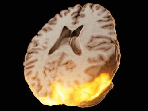
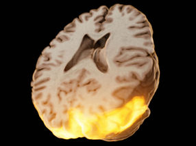
A fusion of anatomical and functional MR imaging, shows the brain areas which are activated during looking at a house.
Fraunhofer MEVIS
Other news from the department science


Get the analytics and lab tech industry in your inbox
From now on, don't miss a thing: Our newsletter for analytics and lab technology brings you up to date every Tuesday. The latest industry news, product highlights and innovations - compact and easy to understand in your inbox. Researched by us so you don't have to.