A New Strategy to Analyze The Cellular World
Scientists develop new tool to analyze cellular structures via high- resolution imaging
In the past two decades, super-resolution microscopy has been one of the fastest evolving fields through many technical improvements. However, the development of new labeling tools, probes and their biological application, is mostly lagging behind the technical capabilities. Most recently, Prof. Silvio O. Rizzoli from the Cluster of Excellence and DFG- Research Center for Nanoscale Microscopy and Molecular Physiology of the Brain (CNMPB) has developed together with his team a new technique that expands the benefit of super- resolution microscopy to study biological questions. This method contributes to understand on how cells renew, distribute and transport their molecular and subcellular components. The new technique was published on May 26th in the Journal of cell biology.
All cells rely on the recycling of membranes via various pathways (secretion, uptake, and membrane turnover). Several types of cellular organelles such as the plasma membrane, the endoplasmic reticulum, the Golgi apparatus, endosomes and vesicles are involved in these processes. However, it was difficult to identify the protein composition of the involved organelles since both, the membranes and the proteins of the same organelle need to be marked simultaneously. Here the main difficulty comes with the membrane probe, as almost all dyes that work excellent in live cell experiments are only poorly fixable and get “lost” during the antibody staining procedure.
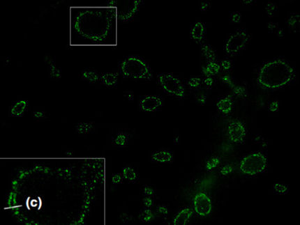
The research team with first author Natalia Revelo therefore developed a membrane probe that overcomes this problem. The probe mCLING (membrane-binding fluorophore-Cysteine- Lysine-Palmitoyl Group) is a composition of a short polypeptide coupled to a membrane anchor and a fluorophore. The study, recently published in the Journal of Cell Biology, shows that mCLING can be used to label the plasma membrane, and also to faithfully track specific organelles, which can be done in conjunction with fixation and immunostaining, in both cell culture and in tissue. The utility of the mCLING probe could be characterized for various important biological model systems and already enabled the authors to answer long-lastin question in the field of membrane recycling. Moreover, mCLING imaging could also be extended to different processes. For example, the structure and molecular organization of isolated organelles in vitro, or the arrangement of proteins on the membranes of various types of cells, can be easily tackled with mCLING. These efforts will be aided by the fact that mCLING can be optimized for any available super-resolution technique.
Original publication
Revelo NH, Kamin D, Truckenbrodt S, Wong AB, Reuter K, Reisinger E, Moser T, Rizzoli SO (2014) A new probe for super-resolution imaging of membranes elucidates trafficking pathways. J CELL BIOL, 205(4): 591-606.
Other news from the department science


Get the analytics and lab tech industry in your inbox
From now on, don't miss a thing: Our newsletter for analytics and lab technology brings you up to date every Tuesday. The latest industry news, product highlights and innovations - compact and easy to understand in your inbox. Researched by us so you don't have to.