Researchers use light to detect Alzheimer's
New technique may help identify ways to predict and prevent deadly disease
A team of researchers in Bedford, Mass. has developed a way of examining brain tissue with near-infrared light to detect signs of Alzheimer's disease. In the March 15 issue of the journal optics Letters, published by the Optical Society of America, the team describes how they used optical technology to examine tissue samples taken from different autopsies and correctly identified which samples came from people who had Alzheimer's disease.
"We're primarily interested in finding a way of diagnosing and monitoring Alzheimer's disease during life," says U.S. Department of Veterans Affairs Research Scientist Eugene Hanlon. "We think this technique has a lot of potential for detecting the disease early on."
The new technique developed by Hanlon and his collaborators at Harvard Medical School/Beth Israel Deaconess Medical Center and Boston University can detect alterations to the optical properties of the brain that occur as the tissue undergoes microscopic changes due to Alzheimer's - sometimes far in advance of clinical symptoms. The technique is now being tested for its effectiveness at diagnosing Alzheimer's disease in living people.
For several years, Hanlon and his colleagues have looked at the possibility of analyzing the brain with near-infrared light, which has the advantage of being able to safely penetrate the skull and pass harmlessly through the brain. Inside the head, some of the infrared light scatters, however, and how the light scatters can tell researchers about the condition of the brain.
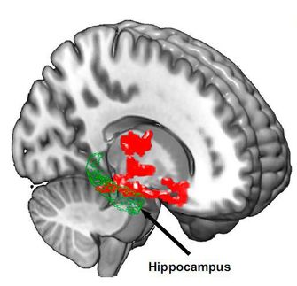
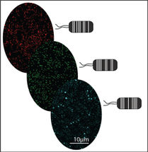
In their paper, the team reports observing an optical effect due to the presence of microscopic features of Alzheimer's. Amyloid plaques, one of the telltale signs of Alzheimer's disease, scatter light differently from normal brain tissue. What Hanlon and his colleagues showed was that as the microscopic plaques accumulate, the optical properties of the brain change. The team found that this change is detectable and that their technique could quantify differences between in-vitro samples and correctly identify signs of Alzheimer's.
This technique will be a boon to medicine if it is able to detect microscopic changes that can be related to disease progression. While techniques like MRI are good at identifying the gross anatomical features associated with Alzheimer's, they cannot detect more microscopic changes.
Original publication: Eugene B. Hanlon et al.; "Scattering Differentiates Alzheimer Disease In Vitro"; Optics Letters 2008, Vol. 33, No. 6.
Other news from the department science


Get the analytics and lab tech industry in your inbox
From now on, don't miss a thing: Our newsletter for analytics and lab technology brings you up to date every Tuesday. The latest industry news, product highlights and innovations - compact and easy to understand in your inbox. Researched by us so you don't have to.