A noninvasive way to view insulin in pancreas
"Scientists and doctors have wanted to know how much insulin a person has in their body, but haven't been able to know the exact amount without the patient being deceased and actually removing the pancreas," says Peter Arvan, division chief of Metabolism, Endocrinology & diabetes at the University of Michigan.
But a new study by Arvan and his fellow researchers finally allowed them to see exactly how much insulin was present in the pancreas of a living animal.
"In the past, there hasn't been technology that would afford us this same opportunity," Arvan says.
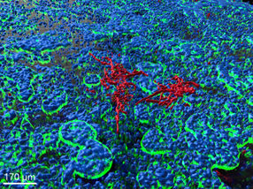
The researchers genetically engineered a mouse that makes the protein that the body uses to produce human insulin, called proinsulin. By engineering the protein as a fluorescent protein, it reacted in a certain way when the researchers shined a fluorescent light on the mouse pancreas. The pancreatic islets -- tiny clusters of cells in the pancreas that include the insulin-making beta cells -- bounced a special wavelength of light back. The strength of the fluorescent signal indicated how much insulin was present.
Researchers were then able to see, in real time, when the mouse was releasing its insulin by giving the mouse sugar water (glucose). After the glucose entered the stomach and blood sugar began to rise, insulin was released in response and fluorescence was released from some of the islets.
When isolated pancreatic beta cells were stimulated the same way, individual fluorescent flashes indicating secretion of small packets of insulin from the cells could be observed (see attached video). Each flash is thought to release between 100,000 and 1 million insulin molecules. When the beta cells were not stimulated, insulin release was very low and no flashes were seen, indicating the amount of fluorescence released from beta cells was proportional to the amount of insulin released.
"We could see responses in specific pancreatic islets that were releasing insulin into the bloodstream," Arvan says. "Amazingly, only a small fraction of the pancreatic islets showed a major response to glucose, while the rest of the islets appeared to remain mostly inactive."
This surprising result implies that some islets represent the body's first line of defense against high blood sugar -- an observation that before this study has never been made.
"Perhaps only after we have lost our first responder islets do we recruit backup islets to provide insulin," Arvan says.
Arvan adds that the study has potential clinical implications that could help patients with diabetes.
"The fact that not all islets are created equal may provide some hope in the future of activating these backup islets to fight diabetes," Arvan says.
"It has been a holy grail to try to look inside a living person's pancreas and see how much insulin that person has stored away," Arvan adds. "This study is an important and exciting first step in understanding how much pancreatic insulin can be produced and secreted in the clinical setting."
Original publication

Other news from the department science


Get the analytics and lab tech industry in your inbox
From now on, don't miss a thing: Our newsletter for analytics and lab technology brings you up to date every Tuesday. The latest industry news, product highlights and innovations - compact and easy to understand in your inbox. Researched by us so you don't have to.
Most read news


More news from our other portals


See the theme worlds for related content
Topic world Fluorescence microscopy
Fluorescence microscopy has revolutionized life sciences, biotechnology and pharmaceuticals. With its ability to visualize specific molecules and structures in cells and tissues through fluorescent markers, it offers unique insights at the molecular and cellular level. With its high sensitivity and resolution, fluorescence microscopy facilitates the understanding of complex biological processes and drives innovation in therapy and diagnostics.