Measurable for the first time: How bio molecules react to lack of space
Sensor demonstrates lack of space in living cells
proteins and other bio molecules are often analysed exclusively in aqueous solutions in test tubes. But it is uncertain if these experimental studies can be transferred to the densely-packed cellular environment. The Bochum-based researchers from the team of Junior Professor Dr Simon Ebbinghaus have developed a novel method which can be used to analyse the effects of the lack of space in living cells with the aid of a microscope for the first time. They designed a sensor that changes colour depending on how confined the space in the cell is. The researchers report their findings in the trade journal Angewandte Chemie and Angewandte Chemie International Edition. The findings are the result of a collaboration conducted between RUB and Max Planck Institute for Coal Research (Dr Matthias Heyden) under the umbrella of the Cluster of Excellence RESOLV.
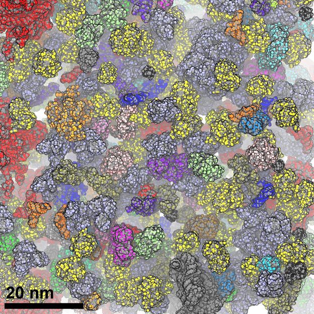
Schematic diagram of the densely-packed cellular environment the compression effects of which are analysed using the introduced sensor.
© RUB, Bild: Physikalische Chemie II
Densely-packed cellular environment
Proteins fulfil a wealth of functions in a cell. Different proteins are responsible for structure formation, catalysis of chemical reactions and transmission of cellular signals. Even the smallest protein defects can have grave consequences: they do, for example, trigger diseases such as Alzheimer’s, Parkinson’s or Huntington’s disease. Therefore, proteins are of much interest for the biochemical and medical research. However, tor the purpose of studies, they are frequently isolated from their natural environment and are analysed in aqueous solutions. “It is often neglected that the cell environment is a densely-packed matrix consisting of diverse macromolecules as well as small organic and inorganic substances, which make the cell interior very viscous and highly concentrated,” explains Simon Ebbinghaus. “This raises the question if analytic methods in aqueous solutions can truly reflect the natural behaviour of proteins in the cellular environment.”
Theory: lack of space in the cell causes compaction of biomolecules
The most common theory for describing the effects of cellular environment is the “excluded volume theory”. Simply put: during the Christmas season, when the Christmas markets, department stores and public transport are crowded, everybody attempts to avoid any contact with the strangers next to them by assuming as compact a posture as possible. This form of repulsion can be translated to proteins, which accordingly assume a compact structure in a densely-packed environment. Because the more compact the proteins, the more stable they are, the theory thus predicts greater stability inside the cell.
Sensor emits light in different colours
In order to better understand these effects, RUB researchers have developed a method for tracking the compression of a macromolecule in a cell. A polymer marked with dyes fulfils the function of a sensor. Once it is compressed in the confined space, the dye pigments move closer together, and the frequency of the measured light changes under the microscope. “Observing the light, a change from green to red can be detected,” describes Simon Ebbinghaus.
Surprise in the living cell
Deploying different macromolecular additives (crowding reagents), which had previously been used to create a stipulated lack of space, RUB researchers successfully demonstrated in the test tube that the sensor is fully functional and that it reacts very sensitively to a densely-packed environment. Subsequently, they injected the sensor into living cells and were surprised: contrary to the expectations, it became apparent that a force exists inside the cell that pulls its compact form apart. This is evidence of attractive forces inside the cell which override the compression effects predicted in theory.
Cell under stress: proteins and functions change
The researchers then put the cell under so-called osmotic stress by increasing the salt concentration in the environment. The cell reacted by releasing water – the result was in an even greater density inside. Such osmotic stress increased the cellular compression effects significantly. “Such changes to the cellular environment can have drastic consequences and affect the protein functions in the long term,” explains Simon Ebbinghaus. “Other forms of stress – for example through unfolded or misfolded proteins which amass in a certain region of the cell – could change the cellular environment significantly. Such changes may promote protein clumping, which play a crucial role in the generation of neurodegenerative diseases.”
Original publication
Other news from the department science


Get the analytics and lab tech industry in your inbox
From now on, don't miss a thing: Our newsletter for analytics and lab technology brings you up to date every Tuesday. The latest industry news, product highlights and innovations - compact and easy to understand in your inbox. Researched by us so you don't have to.