The Combined Nanoscopy Technique
Scientists develop a combined technique for studying cellular structures via high-resolution imaging
Molecular processes in living cells can best be monitored by high-resolution microscopy techniques. Although groundbreaking technical innovations in the field of microscopy have been made in the past, frontiers still exist. Prof. Dr. Silvio O. Rizzoli and his team of the Göttingen DFG Research Center and Cluster of Excellence for Nanoscale Microscopy and Molecular Physiology of the Brains (CNMPB) have now developed a new application by combining two imaging techniques to expand the benefits of high-resolution to study biological questions. The new imaging technique COIN enables to study the turnover and metabolism of subcellular structures, such as organelles, in detail. The new method has been described in Nature Communications.
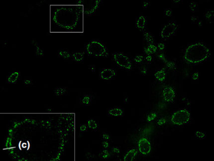
The turnover of subcellular organelles is one of the least understood aspects of modern cell biology, despite its widely recognized importance. In biology, these processes are studied by “feeding” cells with marker molecules such as amino acids labeled with stable isotopes. Over time these amino acids are metabolically incorporated into cellular proteins and the isotopic composition can then be imaged by secondary ion mass spectrometry (SIMS). This technique enables visualization of different organelles in cells and tissues. However, SIMS by itself cannot identify specific subcellular structures.
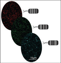
Therefore, the team of Prof. Rizzoli in collaboration with scientists of the Leibniz Institute for Baltic Sea Research in Warnemünde and the French company Cameca successfully correlated SIMS with a second technique. The combined method termed “correlated optical and isotopic nanoscopy (COIN)” is based on super-resolution stimulated emission depletion (STED) microscopy. COIN allows precise studies of the protein turnover in different single organelles from cultured hippocampal neurons. The new approach has been recently published in Nature Communications.
Each of the combined techniques alone provides a piece of information that is unavailable for the other: “SIMS yields the isotopic composition of the material investigated and even its turnover, while STED microscopy reveals the identities and the spatial distribution of organelles or organelle components.”, Prof. Rizzoli explains. The combination (COIN) for the first time allows precisely determining the turnover of proteins in various single organelles in cells. A special feature of the technique is the wide-range application to a variety of biological samples, which should therefore enable the investigation of the composition of many organelles and sub-cellular structures. Using COIN the scientists successfully yielded information about the protein turnover in different organelles of cultured hippocampal neurons. COIN can be applied to a variety of biological samples, and should therefore enable the investigation of the isotopic composition of many organelles and subcellular structures.
Original publication
Saka SK, Vogts A, Kröhnert K, Hillion F, Rizzoli SO*, Wessels J* (2014) Correlated optical and isotopic nanoscopy. NAT COMMUN, 5: 3664.
Other news from the department science


Get the analytics and lab tech industry in your inbox
From now on, don't miss a thing: Our newsletter for analytics and lab technology brings you up to date every Tuesday. The latest industry news, product highlights and innovations - compact and easy to understand in your inbox. Researched by us so you don't have to.
Most read news


More news from our other portals


See the theme worlds for related content
Topic world Fluorescence microscopy
Fluorescence microscopy has revolutionized life sciences, biotechnology and pharmaceuticals. With its ability to visualize specific molecules and structures in cells and tissues through fluorescent markers, it offers unique insights at the molecular and cellular level. With its high sensitivity and resolution, fluorescence microscopy facilitates the understanding of complex biological processes and drives innovation in therapy and diagnostics.
Topic World Mass Spectrometry
Mass spectrometry enables us to detect and identify molecules and reveal their structure. Whether in chemistry, biochemistry or forensics - mass spectrometry opens up unexpected insights into the composition of our world. Immerse yourself in the fascinating world of mass spectrometry!