A deep look into the body with Raman spectroscopy
Raman spectroscopy has recently undergone major advances in the area of deep non-invasive characterisation of biological tissues. The progress stems from the development of spatially offset Raman spectroscopy (SORS) and renaissance of transmission Raman spectroscopy permitting the assessment of diffusely scattering samples at depths several orders of magnitude deeper than possible with conventional Raman spectroscopy.
In a review article, Pavel Matousek (STFC Rutherford Appleton Laboratory, Harwell Oxford) and Nicholas Stone (University of Exeter) explain recent developments within the medical area and give some examples of emerging applications including non-invasive diagnosis of bone disease, cancer and monitoring of glucose levels.
Raman spectroscopy has proven to be an effective technique for analyzing excised bone matrix but the difficulty in probing deep through soft tissue hampered in vivo studies. The development of deep sub-surface Raman techniques provides new opportunities offering promise to diagnose bone conditions, such as brittle bone disease and osteoporosis, noninvasively. For example, the development of a Raman instrument intended for transcutaneous monitoring of steroid-induced osteoporosis has been reported.
A fast developing area for SORS and transmission Raman spectroscopy is presently the non-invasive detection of deep cancer lesions in breast tissue. In a study, it was shown with Raman spectroscopy that the apatite carbonate levels associated with benign and malignant calcifications are significantly different. At present, there is no reliable way to distinguish between the different calcification types by mammography. In studies on tissue phantoms using the Raman Kerr gated, SORS and transmission Raman methods, respectively, recognizable chemical signatures were obtained for the two different calcification types. Recent advances could even make it possible to go beyond the discrimination purely of Types I and II calcifications, to the compositional profiling of the Type II calcifications and the associated disease state, in situ in bulk breast tissue. Yet, further work is still required to reach the top range of clinically relevant tissue depths.
It is often crucial to remove the just right amount of tissue ensuring that no cancerous cells are left within a patient’s body whilst ensuring the amount of tissue removed is kept to the minimum. Recently, the capability of SORS to identify soft tissue lesions at depths of several mm’s within healthy soft tissue was demonstrated. The study heralds prospect for a real time diagnostic tool that could be deployed during surgery to aid surgeons in defining the margins of lesions.
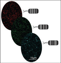
The SESORS concept combining SERS and SORS techniques has been successfully demonstrated in the area of glucose detection recently. Using an implanted sensor, the glucose was successfully monitored in the interstitial fluid of rats by SORS at clinically relevant levels.
Other potential future applications of the SESORS concept when used with appropriate nanoparticle labels, such as antibodies for specific disease cellular expressions can include early cancer detection and monitoring of various other conditions. Especially attractive feature of this concept is its inherent ability for multiplexing. It is likely the use of multiplexed SESORS could lead to advances in tumor specific treatment selection and monitoring in real-time. Furthermore the detection and monitoring for the presence of infectious diseases with SESORS or in combination with minimally invasive methods, with needles or insertable probes, to interrogate inner cavities and vessels present another area of potential future use.
Original publication
P. Matousek and N. Stone, Recent advances in the development of Raman spectroscopy for deep non-invasive medical diagnosis, J. Biophotonics 6(1), 7-19 (2013).
Other news from the department science


Get the analytics and lab tech industry in your inbox
From now on, don't miss a thing: Our newsletter for analytics and lab technology brings you up to date every Tuesday. The latest industry news, product highlights and innovations - compact and easy to understand in your inbox. Researched by us so you don't have to.
Most read news


More news from our other portals


See the theme worlds for related content
Topic World Spectroscopy
Investigation with spectroscopy gives us unique insights into the composition and structure of materials. From UV-Vis spectroscopy to infrared and Raman spectroscopy to fluorescence and atomic absorption spectroscopy, spectroscopy offers us a wide range of analytical techniques to precisely characterize substances. Immerse yourself in the fascinating world of spectroscopy!